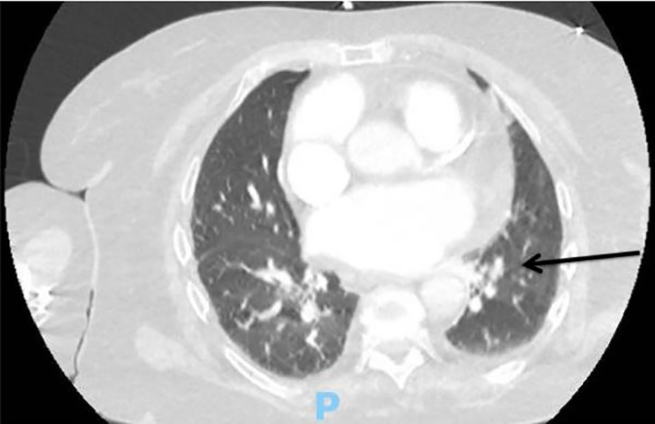
Figure 1. Computed tomography (CT) angiography of the chest upon initial presentation showing a right lower lobe filling defect consistent with segmental pulmonary embolism (black arrow).
| Cardiology Research, ISSN 1923-2829 print, 1923-2837 online, Open Access |
| Article copyright, the authors; Journal compilation copyright, Cardiol Res and Elmer Press Inc |
| Journal website http://www.cardiologyres.org |
Case Report
Volume 10, Number 4, August 2019, pages 249-252
Spontaneous Hemorrhagic Pericardial and Pleural Effusion in a Patient Receiving Apixaban
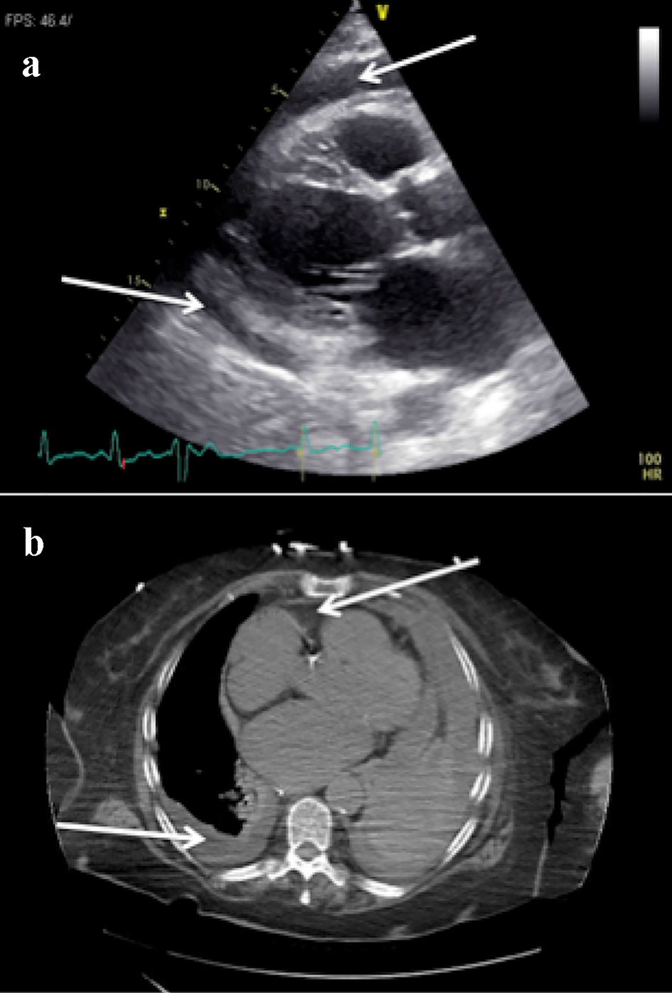
Figures