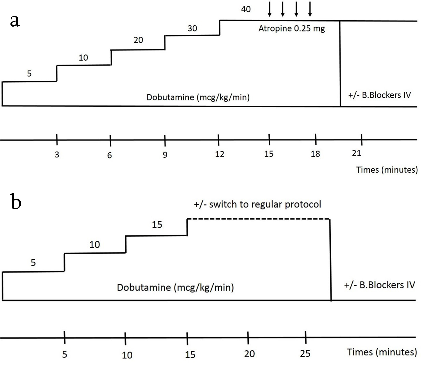
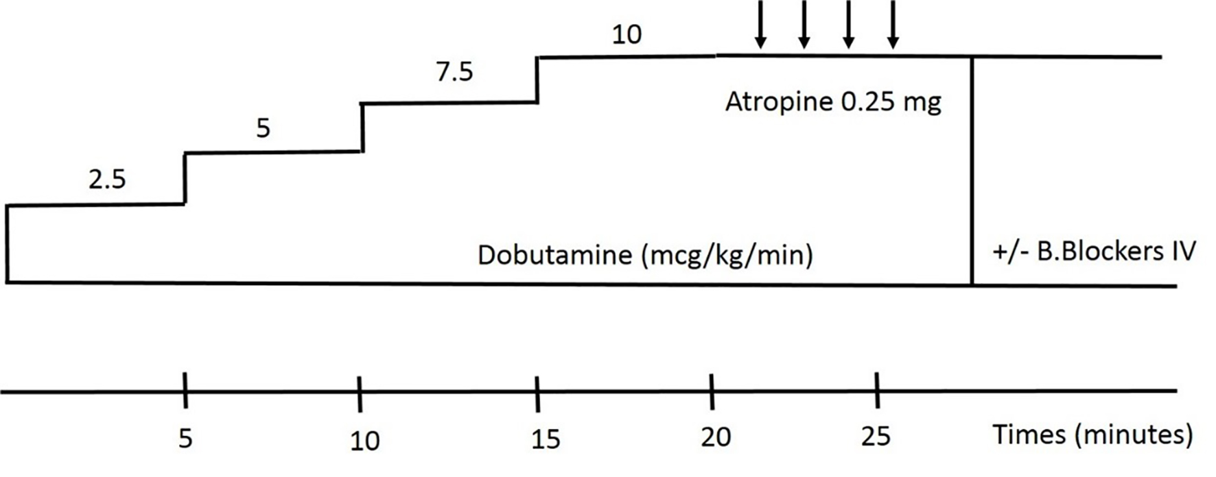
Figure 1. Image showing regular DSE protocol (a) and (b) low dose DSE protocol. DSE: dobutamine stress echocardiography.
| Cardiology Research, ISSN 1923-2829 print, 1923-2837 online, Open Access |
| Article copyright, the authors; Journal compilation copyright, Cardiol Res and Elmer Press Inc |
| Journal website http://www.cardiologyres.org |
Review
Volume 11, Number 2, April 2020, pages 89-96
Stress Echocardiography: Concept and Criteria, Structure and Steps, Obstacles and Outcomes, Focused Update and Review
Figures
Tables
| Rest | Stress | Diagnosis |
|---|---|---|
| Normokinesis | Normo- to hyperkinesis | Normal |
| Normokinesis | Hypokinesis to akinesis or dyskinesis | Ischemia |
| Akinesis | Hypo- to normokinesis | Viable |
| Akinesis or dyskinesis | Akinesis or dyskinesis | Necrosis |
| Nature of tissue | Rest | Low dose dobutamine | Peak dose dobutamine |
|---|---|---|---|
| DSE: dobutamine stress echocardiography. | |||
| Normal segment | Normal | Normal or improves | Hyperkinetic |
| Ischemic segment | Normal | Normal or may worsen | Worsens |
| Viable ischemic | Hypo- or akinetic | Improves | Worsens (biphasic response) |
| Infarcted segment | Akinetic or dyskinetic | Akinetic or dyskinetic | Akinetic or dyskinetic |
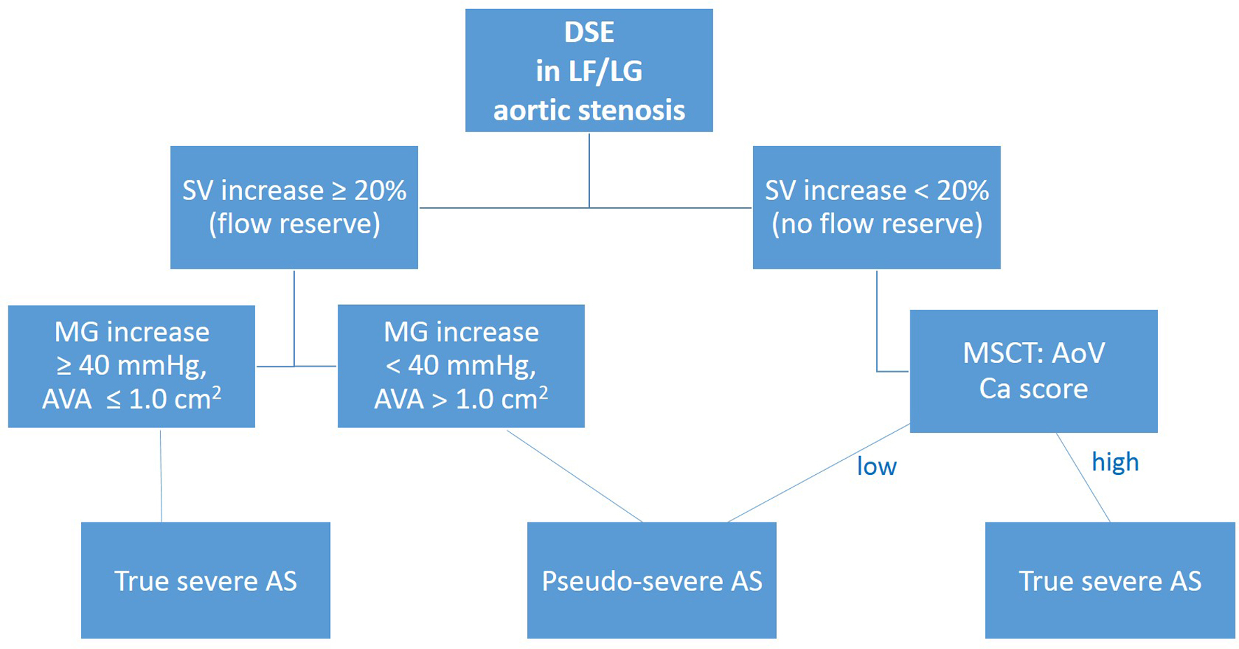
| True severe AS | Pseudo-severe AS | |
|---|---|---|
| +: minor increase; +++: significant increase; =: similar; -: decrease. AS: aortic stenosis; LVOT: left ventricle outflow tract. | ||
| Stroke volume and LVOT velocity | + | + |
| Transvalvular gradients | +++ | + |
| Aortic valve area | = or (-) | + |