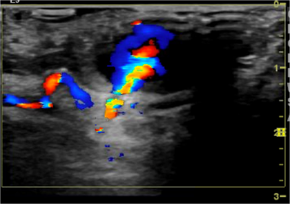
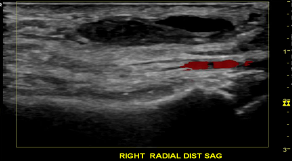
Figure 1. Duplex ultrasound demonstrating patent right radial artery with a pseudoaneurysm just measuring 2.3 cm × 2.4 cm × 1.4 cm, and the neck of the pseudoaneurysm measuring 2.1 mm.
| Cardiology Research, ISSN 1923-2829 print, 1923-2837 online, Open Access |
| Article copyright, the authors; Journal compilation copyright, Cardiol Res and Elmer Press Inc |
| Journal website http://www.cardiologyres.org |
Case Report
Volume 10, Number 2, April 2019, pages 131-134
Noninvasive Treatment Approach of Radial Pseudoaneurysm
Figures
Table
| Time (h) | Inflated air (mL) in compression device | Size of pseudoaneurysm |
|---|---|---|
| 0 | 10 | 2.5 cm |
| 1 | 9 | Not measured |
| 2 | 8 | Not measured |
| 3 | 7 | Not measured |
| 4 | 6 | Not measured |
| 5 | 5 | Not measured |
| 6 | 4 | Not measured |
| 7 | 3 | Not measured |
| 8 | 2 | Not measured |
| 9 | 1 | Not measured |
| 10 | 0 | 0 cm |