Cardiology Research, ISSN 1923-2829 print, 1923-2837 online, Open Access
Article copyright, the authors; Journal compilation copyright, Cardiol Res and Elmer Press Inc
Journal website http://www.cardiologyres.org

Case Report

Volume 10, Number 2, April 2019, pages 114-119


Post Traumatic Pseudoaneurysm of Left Anterior Descending Artery Presenting as Acute Coronary Syndrome

Figures

Figure 1.
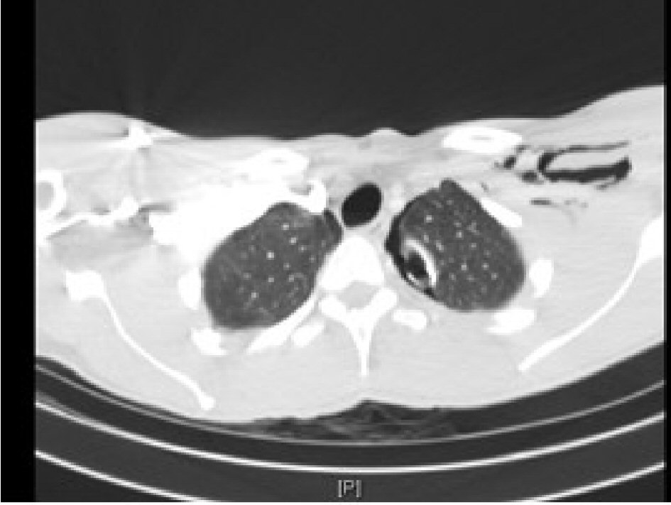
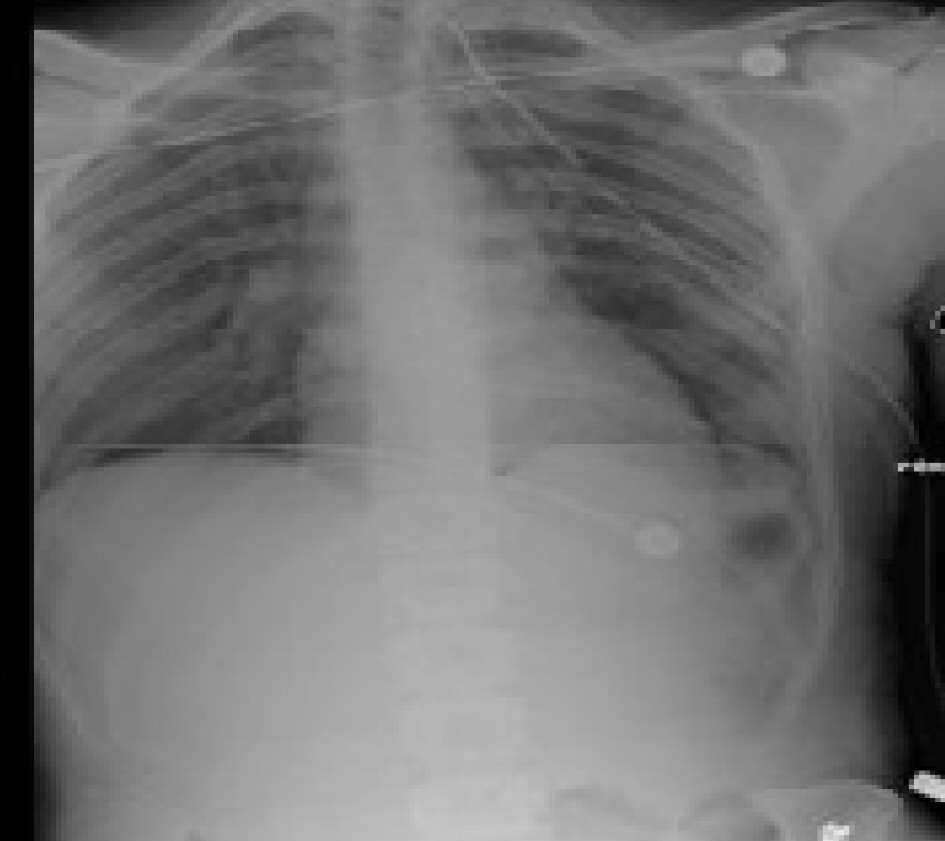
Figure 1. Initial presenting chest X-ray following chest tube insertion.
Figure 2.
Figure 2. CT scan on initial admission demonstrating left-sided subcutaneous air and small residual pneumothorax with chest tube in place.
Figure 3.
Figure 3. Initial EKG on first admission.
Figure 4.
Figure 4. EKG at second admission demonstrating ST elevation.
Figure 5.
Figure 5. Initial cardiac catheterization.
Figure 6.
Figure 6. Intravascular USG showing pseudoaneurysm.
Figure 7.
Figure 7. (a) Coronary angiography showing stenosis. (b) Balloon angioplasty. (c) No stenosis present in the previously balloon angioplasty and stented segment.
Figure 8.
Figure 8. Left coronary angiography 3 days after the second intervention showing no restenosis.