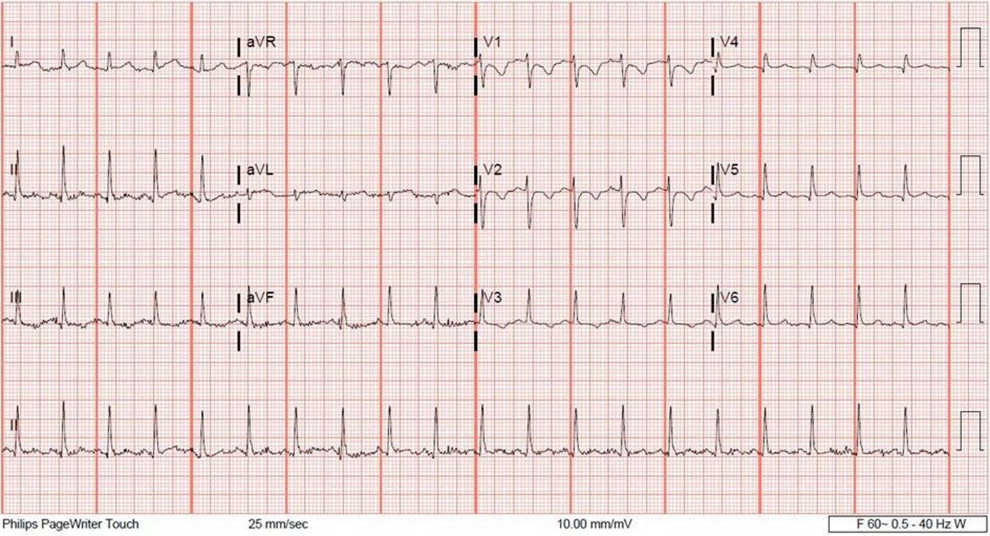
Figure 1. A 12-lead electrocardiogram showing borderline T-wave abnormalities in leads II, III and aVF.
| Cardiology Research, ISSN 1923-2829 print, 1923-2837 online, Open Access |
| Article copyright, the authors; Journal compilation copyright, Cardiol Res and Elmer Press Inc |
| Journal website http://www.cardiologyres.org |
Case Report
Volume 10, Number 1, February 2019, pages 59-62
Mesalamine-Induced Myopericarditis: A Case Report and Literature Review
Figures