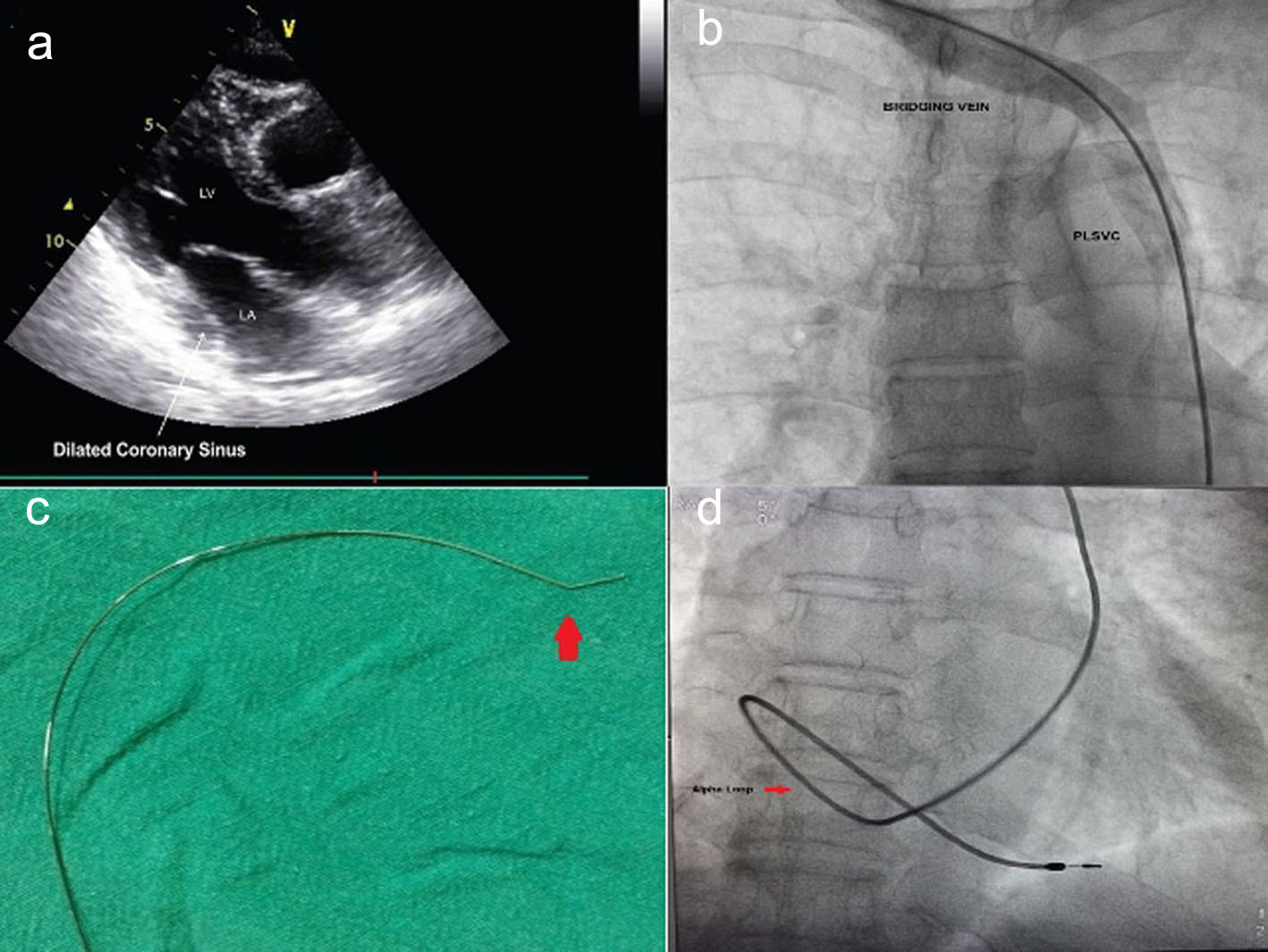
Figure 1. (a) TTE showing dilated coronary sinus. (b) Contrast phlebography from right upper limb showing isolated PLSVC. (c) Mond’s curve. (d) Alpha loop configuration of ventricular lead (passive fixation lead).
| Cardiology Research, ISSN 1923-2829 print, 1923-2837 online, Open Access |
| Article copyright, the authors; Journal compilation copyright, Cardiol Res and Elmer Press Inc |
| Journal website http://www.cardiologyres.org |
Original Article
Volume 10, Number 1, February 2019, pages 18-23
Permanent Pacemaker Implantation in Patients With Isolated Persistent Left Superior Vena Cava From a Right-Sided Approach: Technical Considerations and Follow-Up Outcome
Figures
Tables
| Agitated saline contrast | RSVC | Isolated PLSVC draining to RA | PLSVC with RSVC | LSVC draining into LA |
|---|---|---|---|---|
| LA: left atrium; PLSVC: persistent left superior vena cava; RSVC: right superior vena cava; RA: right atrium; CS: coronary sinus. | ||||
| Contrast from the left arm | RA | CS to RA | CS to RA | LA |
| Contrast from the right arm | RA | CS to RA | RA | RA |
| Baseline characteristics | PM-PLSVC (n = 31, %) | PM-without PLSVC (n = 93, %) | P-value |
|---|---|---|---|
| AV block: atrioventricular block; BFB: bifascicular block; DM: diabetes mellitus; EF: ejection fraction; HTN: hypertension; NYHA: New York Heart Association; PLSVC: persistent left superior vena cava; PM: pacemaker implantation; SND: sinus node dysfunction; TFB: trifascicular block. | |||
| Age (years) | 65.3 ± 11.6 | 64.4 ± 9.3 | 0.6 |
| Sex (men/women) | 19/12 | 57/36 | 0.5 |
| HTN | 6 (19%) | 22 (24%) | 0.4 |
| DM | 5 (16%) | 19 (21%) | 0.23 |
| Ejection fraction (%) | 62.5 ± 4 | 63.6 ± 5 | 0.34 |
| Pacing indication | |||
| SND | 13 (44%) | 42 (45%) | 0.18 |
| AV block | 13 (44%) | 34 (37%) | 0.16 |
| Chronic BFB and TFB | 05 (12%) | 17 (18%) | 0.2 |
| Types of pacemaker | |||
| VVI/VVIR | 09 (29) | 36 (39) | 0.4 |
| DDD/DDDR | 22 (71) | 57 (61) | 0.2 |
| Types of pacing electrode | |||
| Tiened | 05 (16) | 14 (15) | 0.3 |
| Screwing | 26 (84) | 79 (85) | 0.19 |
| Baseline characteristics | PM-PLSVC (n = 31, %) | PM-without PLSVC (n = 93, %) | P-value |
|---|---|---|---|
| PM: pacemaker implantation. | |||
| Procedural time (min) | 25 ± 11 | 23 ± 12 | 0.24 |
| Fluroscopic time (min) | 3.1 ± 2.2 | 2.7 ± 2.1 | 0.54 |
| Length of leads (cm) | |||
| Atrial | 53 | 53 | NS |
| Ventricular | 58 | 58 | NS |
| Pacing parameters | |||
| Atrial lead | |||
| Threshold (mV) | 1.2 ± 0.1 | 1 ± 0.2 | 0.4 |
| P-wave (mV) | 4.1 ± 0.2 | 4.3 ± 0.1 | 0.5 |
| Lead impedance (Ω) | 580 ± 170 | 540 ± 140 | 0.2 |
| Ventricular lead | |||
| Threshold (mV) | 0.8 ± 0.3 | 0.9 ± 0.2 | 0.17 |
| R-wave (mV) | 14 ± 4 | 16 ± 1 | 0.32 |
| Lead impedance (Ω) | 800 ± 240 | 760 ± 20 | 0.22 |
| Alpha loop (ventricular lead) | 31 (100) | 00 | 0.002 |
| Lead dislodgement | 01 (3.2) | 03 (4.8) | 0.32 |
| Subclavian crush | 00 | 00 | 0.00 |
| Local site complication | 02 (6.4) | 07 (7.5) | 0.5 |
| Follow-up duration (years) | 6.9 ± 1.3 | 7.2 ± 1.1 | 0.18 |