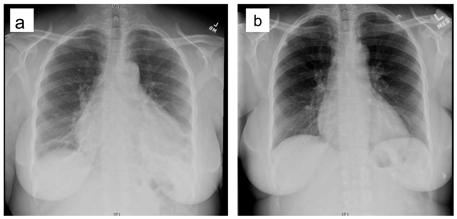
Figure 1. (a) CXR of the patient in current admission. (b) CXR of the patient 5 months prior to this admission.
| Cardiology Research, ISSN 1923-2829 print, 1923-2837 online, Open Access |
| Article copyright, the authors; Journal compilation copyright, Cardiol Res and Elmer Press Inc |
| Journal website https://www.cardiologyres.org |
Case Report
Volume 8, Number 4, 2017, pages 161-164
Chest Pain Due to Pericardial Effusion as Initial Presenting Feature of Rheumatoid Arthritis: Case Report and Review of the Literature
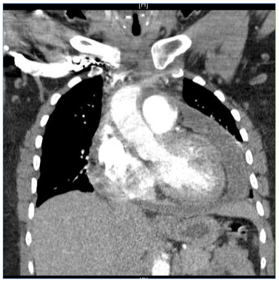
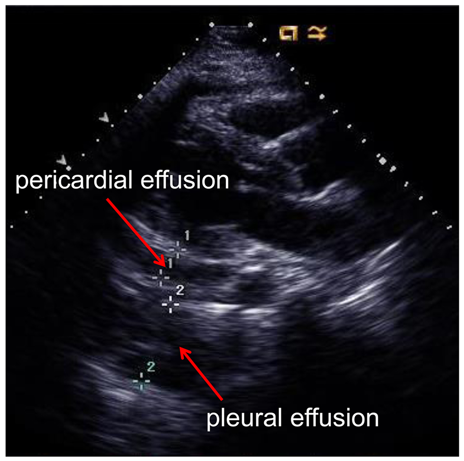
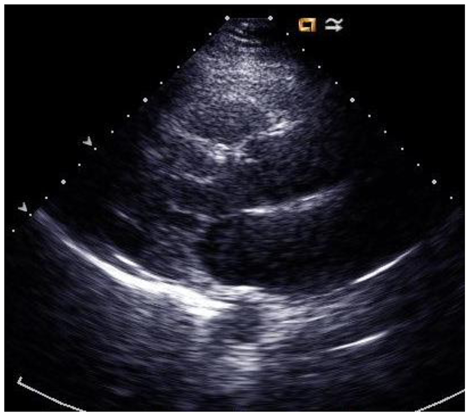
Figures
Table
| Test | RF | Anti-CCP | ANA | Anti-ds-DNA | ESR | CRP |
|---|---|---|---|---|---|---|
| Reference range | Negative | < 20 U | Negative | < 4 IU/mL | 0 - 30 | 0.70 - 1.00 |
| Result | Negative | > 250 U | Negative | < 4 IU/mL | 93 | 8.90 |