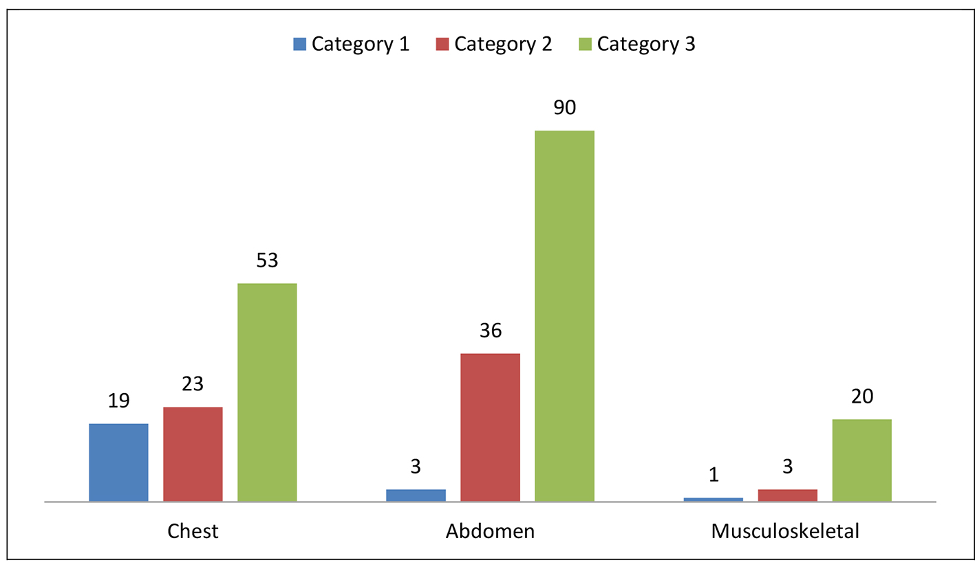
Figure 1. Number and distribution of 248 non-cardiovascular incidental findings in 67 patients by system and categories.
| Cardiology Research, ISSN 1923-2829 print, 1923-2837 online, Open Access |
| Article copyright, the authors; Journal compilation copyright, Cardiol Res and Elmer Press Inc |
| Journal website https://www.cardiologyres.org |
Original Article
Volume 8, Number 1, February 2017, pages 13-19
Non-Cardiovascular Computed Tomography Incidental Findings in Patients Who Underwent Transaortic Valve Implantation Procedure
Figure
Tables
| Total number of patients (n) | 67 |
| Men (n, %) | 35 (52%) |
| Women (n, %) | 32 (48%) |
| Age (years) | 73 ± 8 |
| Body mass index (kg/m2) | 31 ± 7.3 |
| DM (n, %) | 52 (78%) |
| HTN (n, %) | 57 (86%) |
| Hypercholesterolemia (n, %) | 35 (52%) |
| History of smoking (n, %) | 6 (9%) |
| Category | Finding | Number (percentage) |
|---|---|---|
| 1 | Atelectasis and consolidation | 1 (1%) |
| Atelectasis: more than segmental | 1 (1%) | |
| Atelectasis and bronchiectasis | 1 (1%) | |
| Pulmonary edema | 3 (3%) | |
| Moderate to large pleural effusion | 6 (6%) | |
| Pericardial effusion | 3 (3%) | |
| Patchy ground glass opacities | 2 (2%) | |
| Tree in bud with patchy air space opacities | 2 (2%) | |
| 2 | Lung nodules | 10 (11%) |
| Hilar soft tissue with calcification and enactment of the adjacent structures | 1 (1%) | |
| Thyroid nodules | 6 (6%) | |
| Thyroid enlargement | 3 (3%) | |
| Thyroid calcifications | 3 (3%) | |
| 3 | Mosaic attenuation | 18 (19%) |
| RML/lingual sub-segmental atelectasis | 6 (6%) | |
| Emphysema | 2 (2%) | |
| Small pericardial effusion | 1 (1%) | |
| Pleural thickening, likely benign | 1 (1%) | |
| Basal reticulation and GGO | 4 (4%) | |
| Bronchiectasis with no signs of infection | 3 (3%) | |
| Basal fibrotic lung changes | 2 (2%) | |
| Small pleural effusion | 1 (1%) | |
| Calcified mediastinal LN | 3 (3%) | |
| Several small to borderline in size mediastinal lymph node | 9 (9%) | |
| Granuloma | 1 (1%) | |
| Pleural calcification | 2 (2%) | |
| Scattered small cysts | 1 (1%) |
| Category | Finding | Number (percentage) |
|---|---|---|
| 1 | Moderate amount of ascites | 3 (2%) |
| 2 | Prostate enlargement (seven with calcification) | 15 (12%) |
| Adrenal nodule | 4 (5%) | |
| Hypodense liver lesion | 4 (5%) | |
| Hypervascular liver lesion | 3 (2%) | |
| Liver capsular retraction and hypodensity | 1 (1%) | |
| Features of liver cirrhosis | 1 (1%) | |
| Cystic pancreatic lesion | 1 (1%) | |
| Renal cyst with possible internal enhancement | 1 (1%) | |
| Persplenic partly calcified soft tissue mass | 1 (1%) | |
| Fluid filled heterogeneous uterus | 1 (1%) | |
| 3 | Diverticulosis | 14 (11%) |
| Gallstones | 4 (5%) | |
| Splenomegaly with large SV and PV | 1 (1%) | |
| Hiatal hernia | 3 (2%) | |
| Duodenal lipoma | 1 (1%) | |
| Dilated esophagus | 1 (1%) | |
| Absence left kidney | 1 (1%) | |
| Renal stone | 1 (1%) | |
| Simple renal cyst | 26 (20%) | |
| Renal scaring | 2 (2%) | |
| Renal atrophy | 5 (4%) | |
| Renal infarction | 2 (2%) | |
| Fibroid | 6 (5%) | |
| Fatty liver | 14 (11%) | |
| Calcified abdominal lymph nodes | 1 (1%) | |
| Splenic infraction | 2 (2%) | |
| Liver cyst | 1 (1%) | |
| Peripheral splenic calcification | 1 (1%) | |
| Transient intussusception | 1 (1%) | |
| Gastric diverticulum | 1 (1%) |
| Category | Finding | Number (percentage) |
|---|---|---|
| 1 | Anterior abdominal wall thickening, fat stranding with foci of air, query cellulitis | 1 (4%) |
| 2 | Osteopenia with non-aggressive iliac lytic areas | 1 (4%) |
| Lumbar vertebrae complete collapse with posterior bulge | 1 (4%) | |
| Non-aggressive well defined lytic iliac bone lesion with internal calcification | 1 (4%) | |
| 3 | Spondololysis | 3 (13%) |
| Sacroilitots | 1 (4%) | |
| DISH | 1 (4%) | |
| Spine compression fracture | 2 (8%) | |
| Elastofibroma dorsi | 1 (4%) | |
| Non-complicated abdominal wall hernia | 5 (21%) | |
| Gynecomastia | 2 (8%) | |
| Muscle lipoma | 2 (8%) | |
| Breast calcification | 1 (4%) | |
| Mild scoliosis | 1 (4%) | |
| Calcified nodes in the axillary region and pelvic muscles | 1 (4%) |