Figures
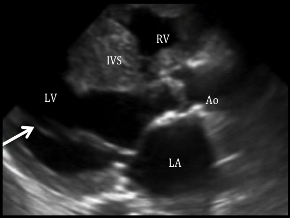
Figure 1. Parachute mitral valve with mitral stenosis on parasternal long axis view. Parasternal long axis view shows both chordae are attached to a single point (arrow), consistent with a parachute mitral valve, giving rise to a mitral stenosis. The left atrium (LA) is dilated with an LA volume index of 45 mL/m2. In this view, the aortic valve seems to be thickened. RV: right ventricle; IVS: interventricular septum; LV: left ventricle; Ao: aorta; LA: left atrium.
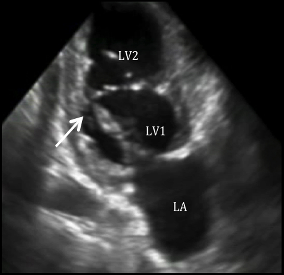
Figure 2. Double chambered left ventricle on two-chamber view. Two-chamber view shows a double chambered LV.
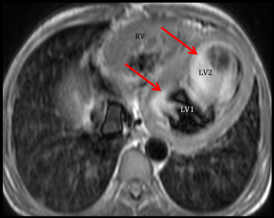
Figure 3. Cardiac MRI demonstrates the double chambered LV. Axial view shows that the LV is partially compartmentalized into two chambers. Note that there is marked flow stasis at the apical part of the LV (LV2) compared to that of the base (LV1). Ejection fraction by cardiac MRI was 15%.
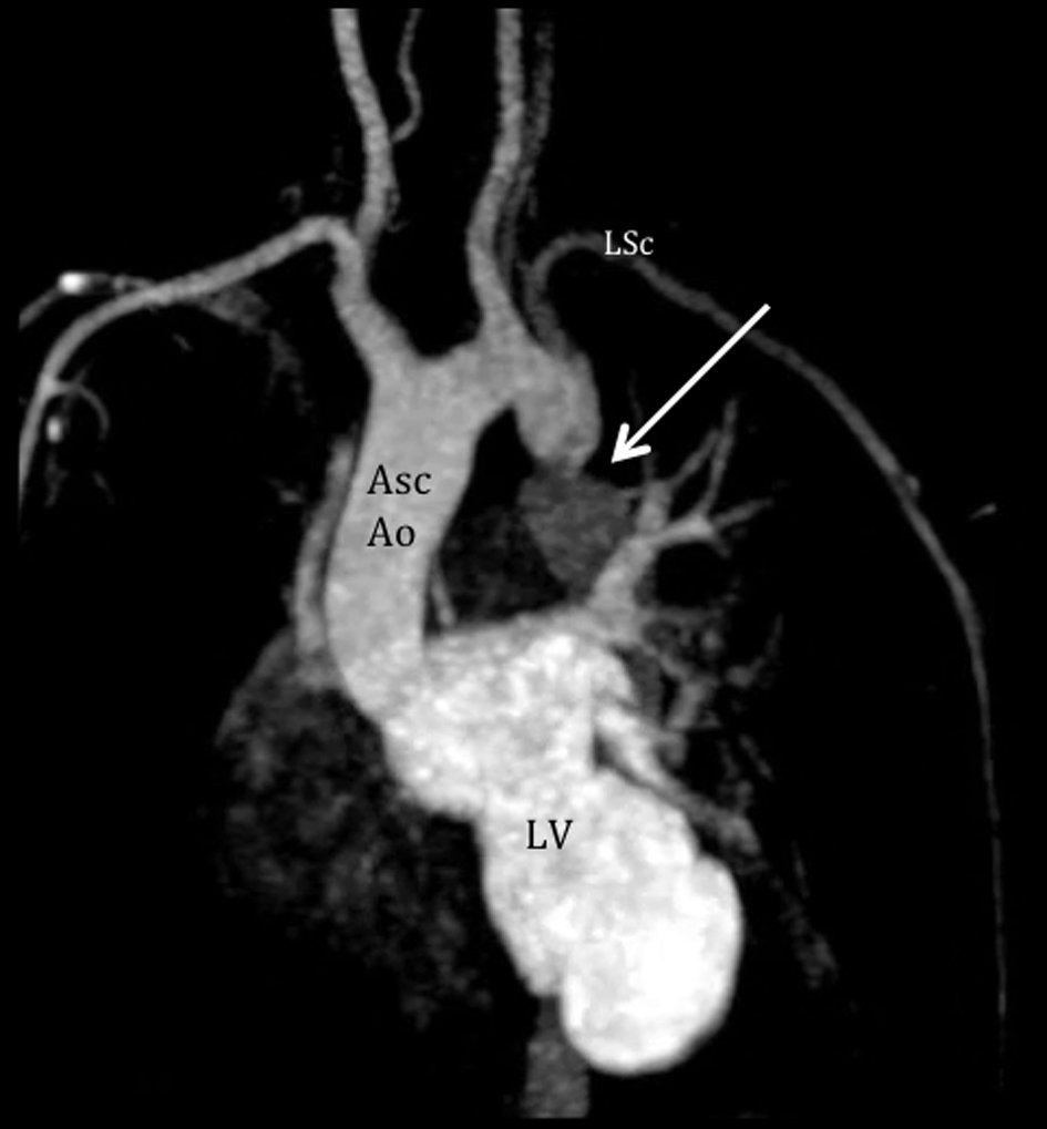
Figure 4. Cardiac MRI demonstrates the coarctation of the aorta. Aortogram shows severe juxtaductal coarctation of the aorta (arrow), 20 mm from the take off of the left subclavian artery (LSc). There is antegrade flow from the LV with significant compromise after the coarctation. Asc Ao: ascending aorta; LSc: left subclavian artery; LV: left ventricle.
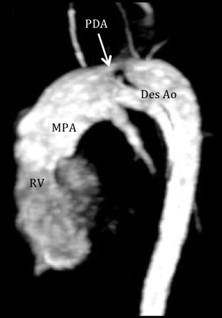
Figure 5. Cardiac MRI demonstrates a patent ductus arteriosus. Pulmonary angiogram shows flow from the pulmonary artery to the descending aorta through a patent ductus (arrow), measuring 8 mm wide and 15 mm in length. RV: right ventricle; MPA: main pulmonary artery; PDA: patent ductus arteriosus; Des Ao: descending aorta.
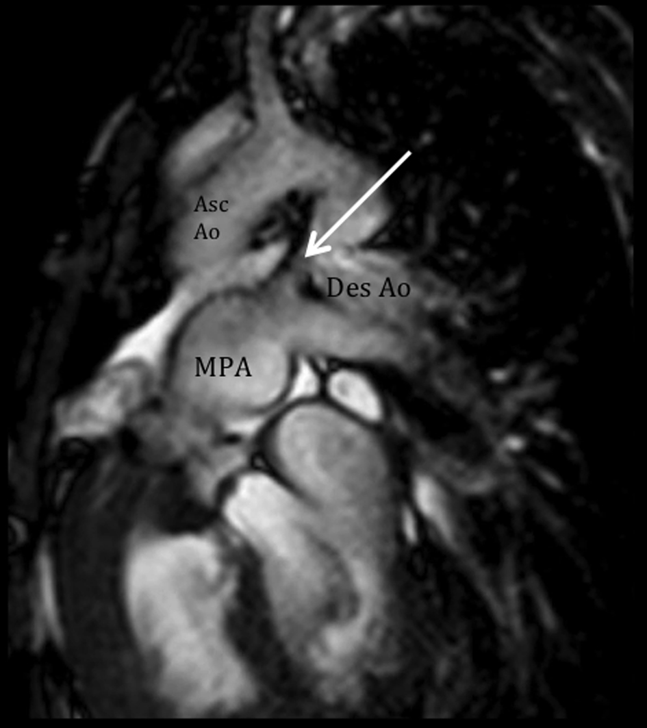
Figure 6. Altered hemodynamics due to multiple anomalies. Cine and velocity flow images show two turbulent jets, consistent with significant right-to-left shunting: 1) an antegrade flow from the dilated pulmonary artery through the ductus (arrow) towards the descending aorta, and 2) a retrograde flow towards the left subclavian artery, predominantly during systole. MPA: main pulmonary artery; Asc Ao: ascending aorta; Des Ao: descending aorta.