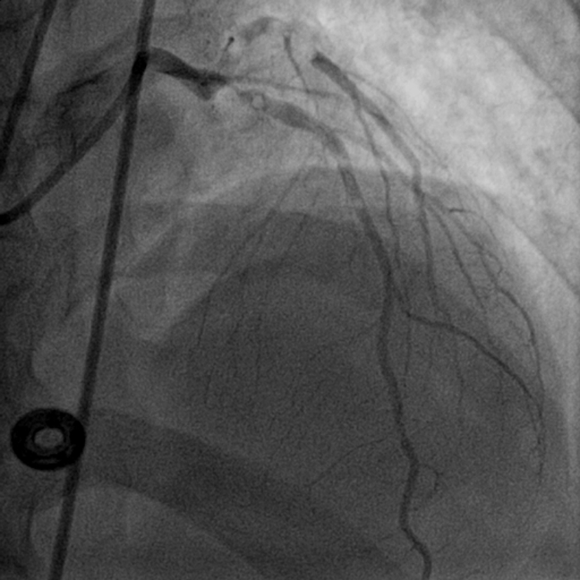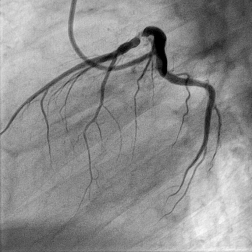
Figure 1. LAO view showing discrete lesion with 90% stenosis of proximal LAD (LAO: left anterior oblique view).
| Cardiology Research, ISSN 1923-2829 print, 1923-2837 online, Open Access |
| Article copyright, the authors; Journal compilation copyright, Cardiol Res and Elmer Press Inc |
| Journal website https://www.cardiologyres.org |
Case Report
Volume 6, Number 1, February 2015, pages 236-238
Massive Coronary Air Embolism Treated Successfully by Simple Aspiration by Guiding Catheter
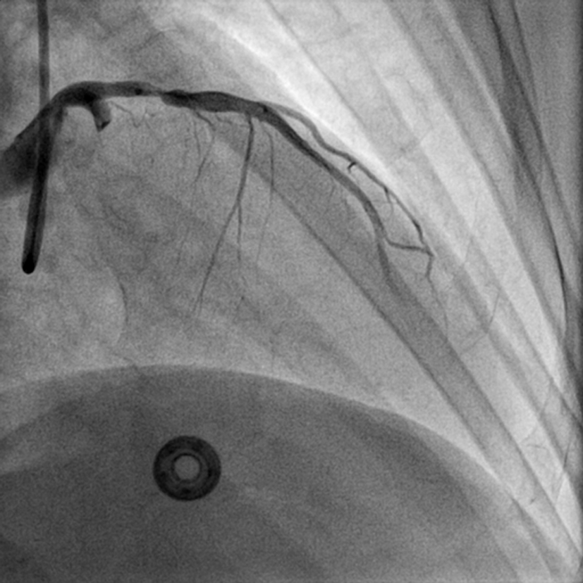
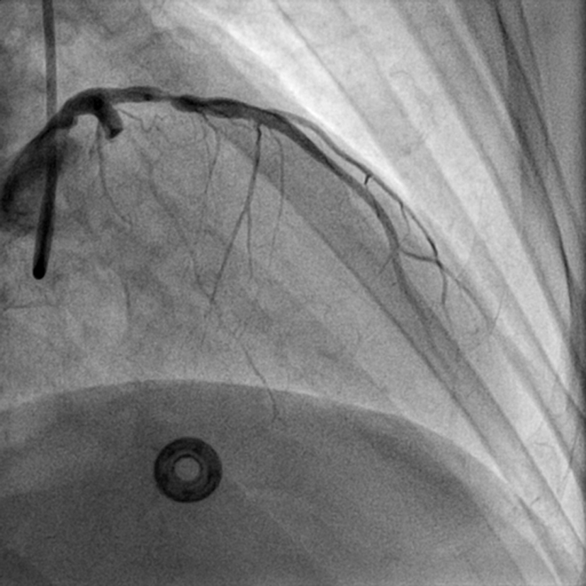
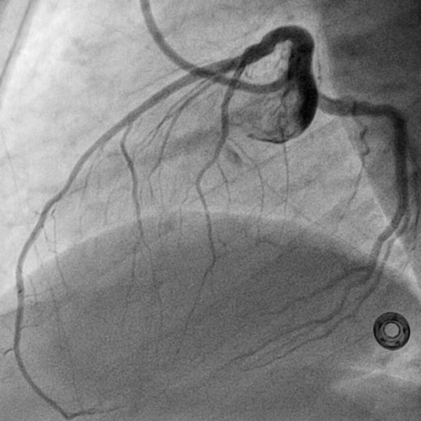
Figures