Cardiology Research, ISSN 1923-2829 print, 1923-2837 online, Open Access
Article copyright, the authors; Journal compilation copyright, Cardiol Res and Elmer Press Inc
Journal website https://www.cardiologyres.org

Review

Volume 14, Number 5, October 2023, pages 319-333


Subvalvular Aortic Stenosis: Learning From Human and Canine Clinical Research

Figures

Figure 1.
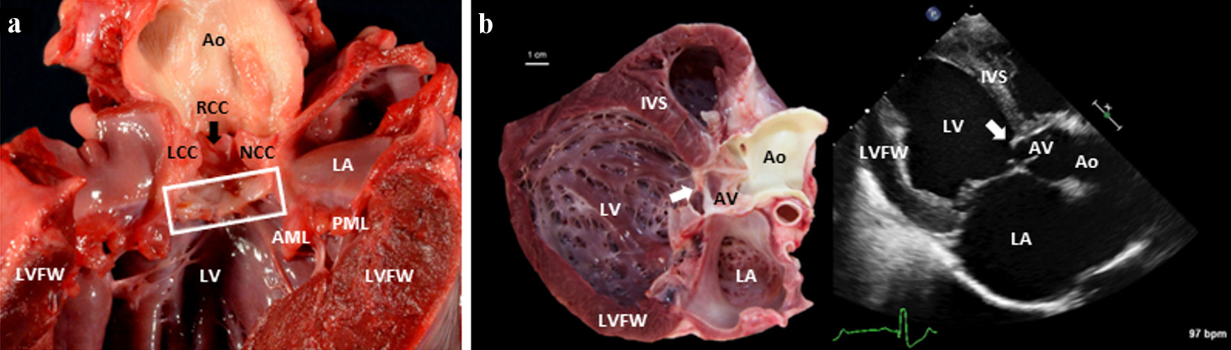
Figure 1. (a) A gross pathology image of the left side of a canine heart affected with SAS. The heart was incised along the long axis of the heart, from the apex to the base (transecting the LVFW). A box surrounds the subvalvular aortic ridge. A black arrow indicates the RCC. (b) A paired gross image and a 2D echocardiographic image (from a right parasternal long-axis five-chamber view) of an 8-month-old Mastiff dog heart afflicted by severe SAS. In both images, you can see the white (gross pathology specimen) and bright (2D echocardiographic image) severe subvalvular ridge/ring of tissue indicated by the white arrow. Note that the specimen shows a dilated and concentrically hypertrophied left ventricle in response to the pressure overload of SAS and the volume overload of concomitant severe AI and mitral valve regurgitation. 2D: two-dimensional; AML: anterior mitral leaflet; Ao: aorta; IVS: interventricular septum; LA: left atrium; LCC: left coronary cusp; LV: left ventricular lumen; LVFW: left ventricular free wall; LVOT: left ventricular outflow tract; NCC: noncoronary cusp; PML: posterior mitral leaflet; RCC: right coronary cusp; SAS: subvalvular aortic stenosis.
Figure 2.
Figure 2. Descriptions and depictions of aortic stenosis in its many forms and the terminology pertinent to this review of subvalvular aortic stenosis. Images are two-dimensional, cross-sectional cartoon illustrations (rotated approximations of right parasternal long-axis five-chamber view). Pathology is illustrated in the context of the human heart. Depictions in a canine heart would be similar, with minor anatomical differences. HOCM: hypertrophic obstructive cardiomyopathy; LVH: left ventricular hypertrophy; LVOT: left ventricular outflow tract; SAM: systolic anterior motion.
Figure 3.
Figure 3. A histopathology sample from a 5-year-old Boerboel with severe SAS stained by hematoxylin and eosin. The open arrow represents an aortic valve leaflet, the double asterisk denotes the aortic wall, the single asterisk is shown within the proximal IVS, and the pound sign denotes the adjacent subvalvular ridge responsible for the increased LVOT pressure gradient. Note the thickening of the aortic valve leaflet in response to turbulent blood flow. IVS: interventricular septum; LVOT: left ventricular outflow tract; SAS: subvalvular aortic stenosis.

Table

Table 1. Comparison Between Canine and Human Disease Characteristics
 
Human subvalvular aortic stenosisCanine subvalvular aortic stenosis
CHD: congenital heart disease; LVOT: left ventricular outflow tract; EOA: effective orifice area; 2D: two-dimensional; 3D: three-dimensional; ECG: electrocardiogram; TXR: thoracic radiographs; TEE: transesophageal echocardiography; MST: median survival time; SAS: subvalvular aortic stenosis; AI: aortic insufficiency.
Prevalence0.25% of children, 6% of CHD cases, and 8-30% of LVOT obstruction cases0.3% of patients, 4.7% of cardiology patients, and 24% of CHD cases
Pattern of inheritanceUnknownLikely autosomal dominant with incomplete penetrance or polygenic
DiagnosisEchocardiogram (EOA, 2D, M-mode, Doppler, 3D, and TEE), ECG, and TXR +/- catheterization and bloodworkEchocardiogram (2D, M-mode, and Doppler) +/- ECG, TXR, and bloodwork
Therapy/interventionResection of fibrous tissue +/- myectomy +/- Konno aortoventriculoplastyAtenolol (a beta-adrenergic blocker)
OutcomesMST is not significantly different from people without SAS; AI, recurrence of fibrous tissue and re-operationMST is 56 months with atenolol; left-sided congestive heart failure, endocarditis, arrhythmias, exercise intolerance, syncope, and sudden death
Environment/exposureRange is similar to nearly identical