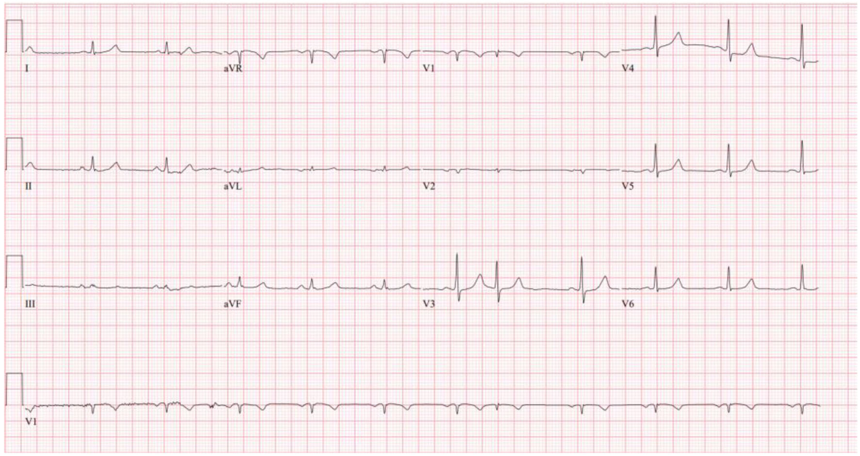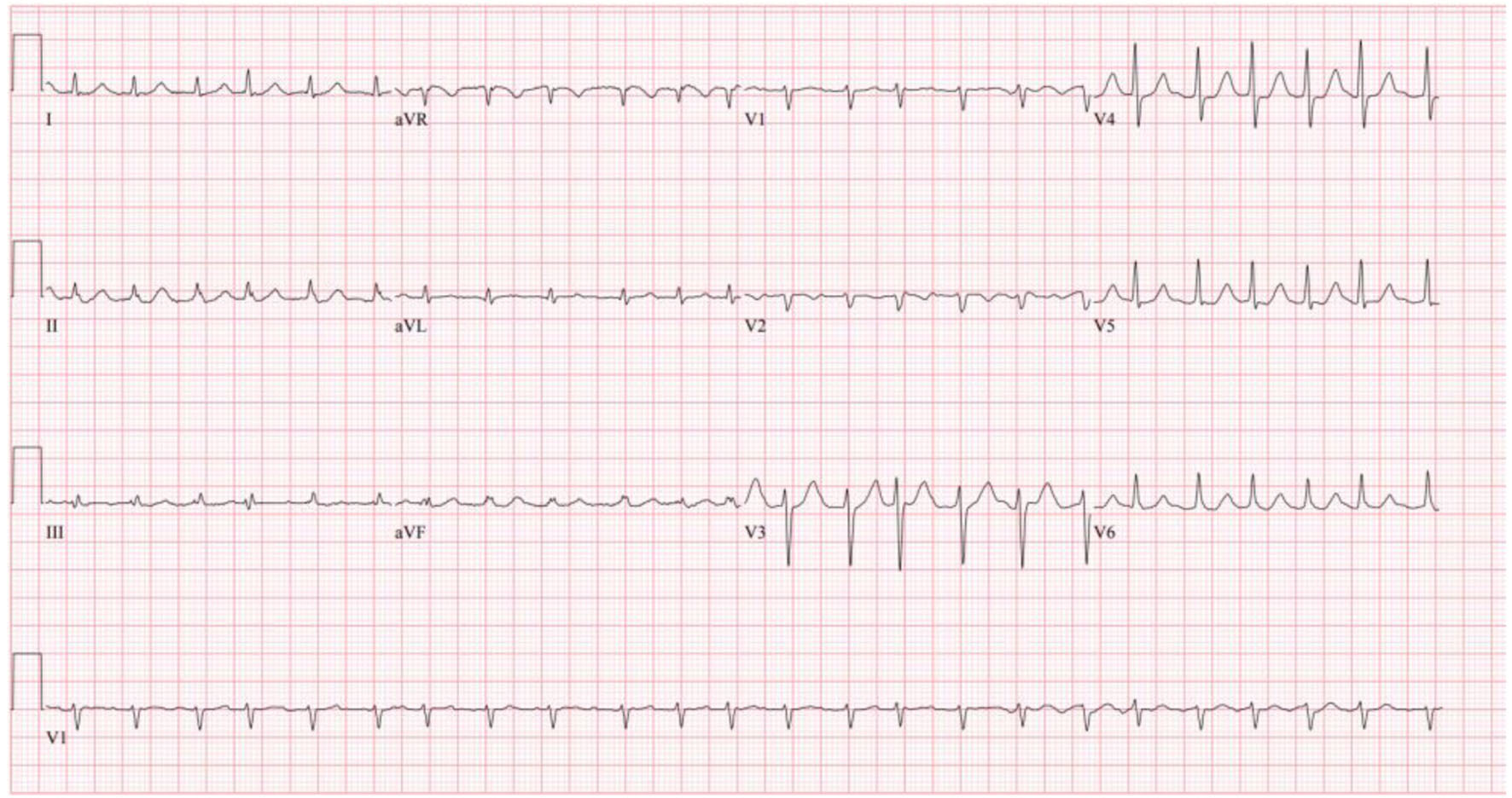
Figure 1. Patient’s initial ECG on admission showing atrial fibrillation with RVR. ECG: electrocardiogram; RVR: rapid ventricular rate.
| Cardiology Research, ISSN 1923-2829 print, 1923-2837 online, Open Access |
| Article copyright, the authors; Journal compilation copyright, Cardiol Res and Elmer Press Inc |
| Journal website https://www.cardiologyres.org |
Case Report
Volume 14, Number 1, February 2023, pages 79-85
An Uncommon Case of Atrial Fibrillation due to a Lung Mass Invasion of the Left Atrial Cavity
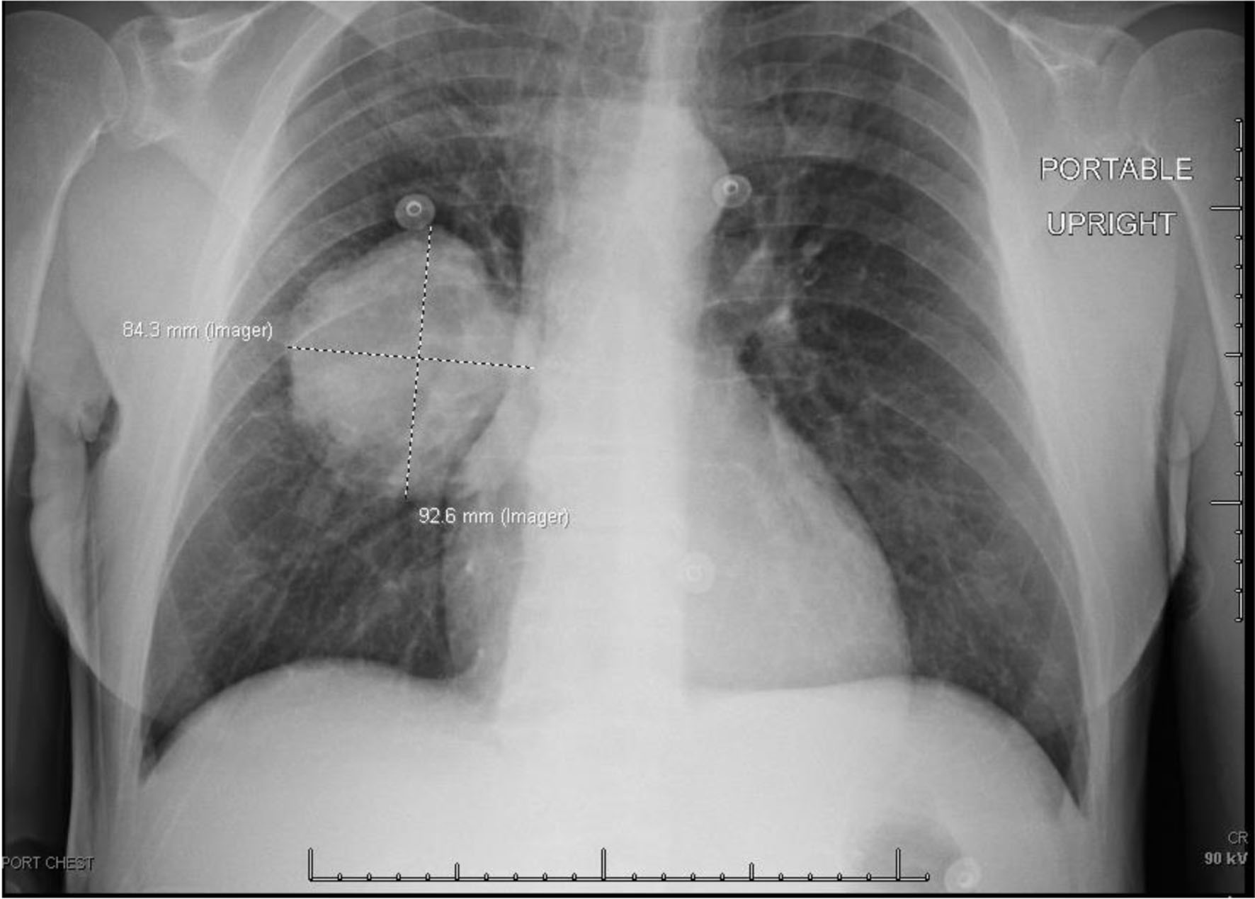
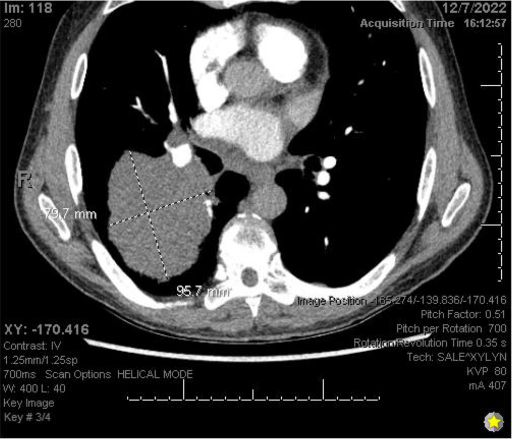
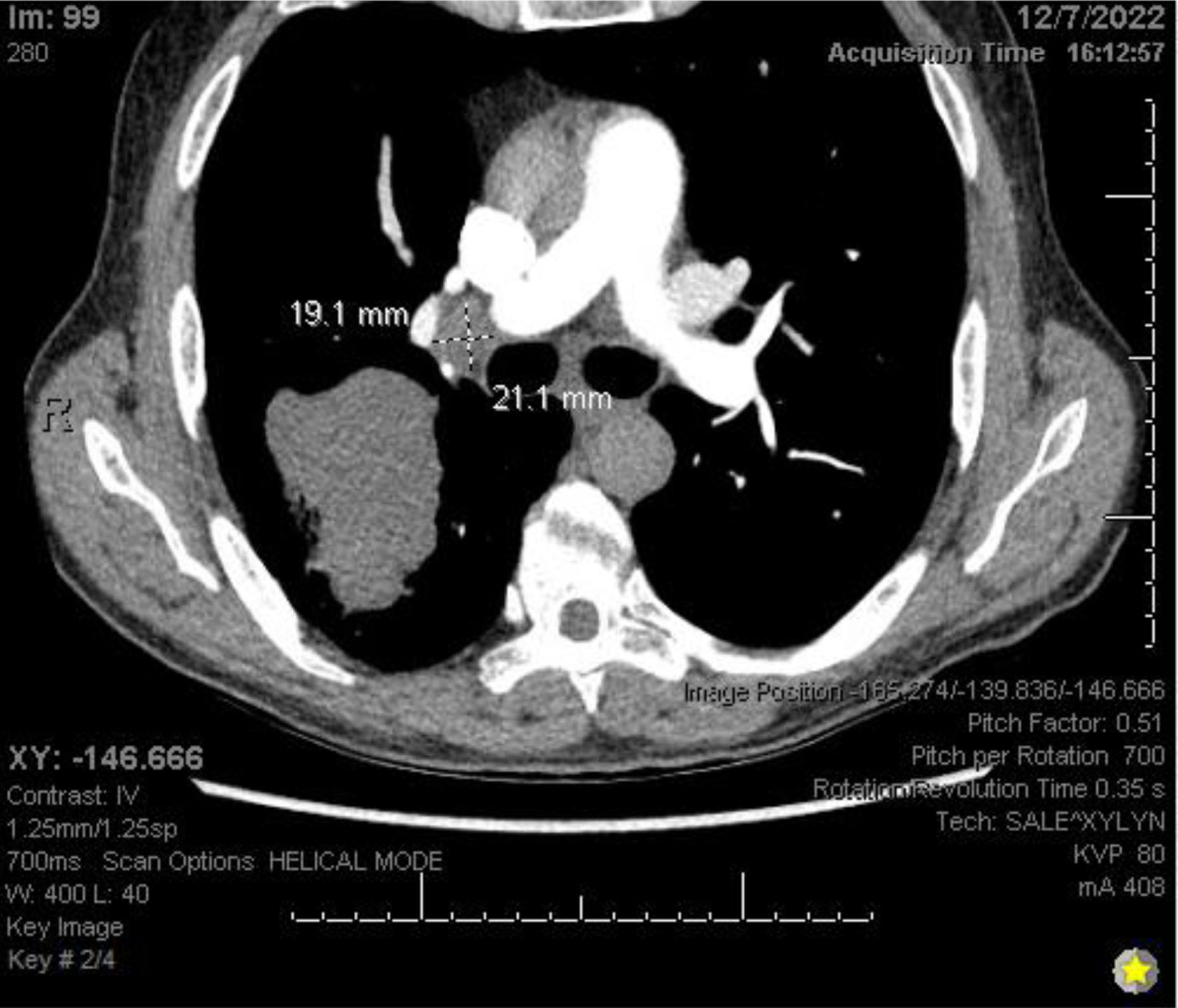
Figures