Cardiology Research, ISSN 1923-2829 print, 1923-2837 online, Open Access
Article copyright, the authors; Journal compilation copyright, Cardiol Res and Elmer Press Inc
Journal website https://www.cardiologyres.org

Case Report

Volume 13, Number 6, December 2022, pages 398-404


Overlapping Phenotype of Adult-Onset ALPK3-Cardiomyopathy in the Setting of Two Novel Variants

Figures

Figure 1.
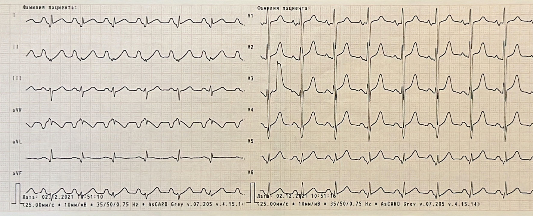
Figure 1. Electrocardiogram showing sinus rhythm, hypertrophy of both atriums and right ventricle, poor progression R in V4 - V6, pathological Q wave in lateral leads, ST segment elevation in I, V5 and V6, low QRS voltage in standard and V5 - V6 leads.
Figure 2.
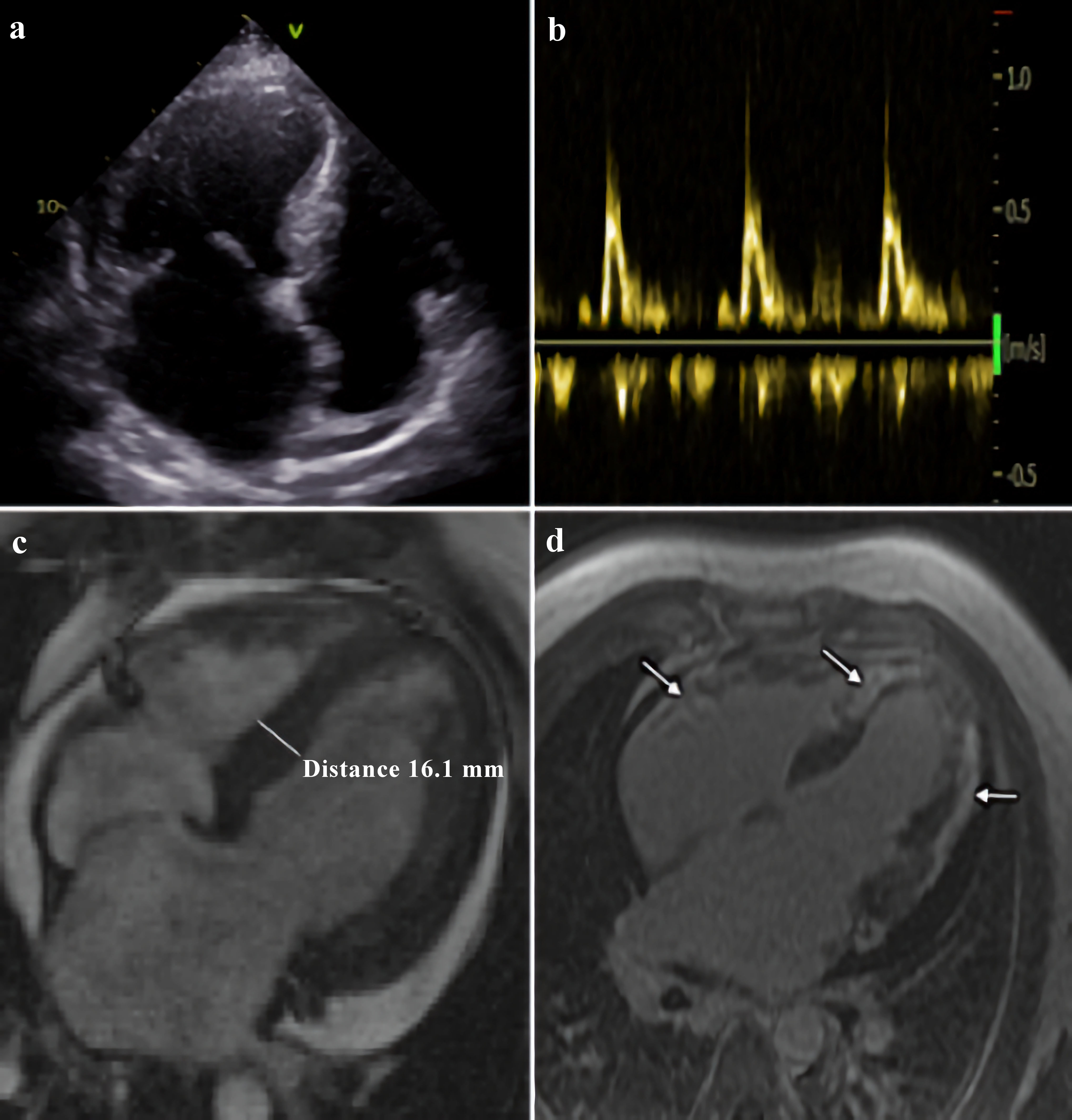
Figure 2. Morphological features of ALPK3-associated cardiomyopathy. Echocardiogram: apical four-chamber view (a), and transmitral flow, demonstrating restrictive type of LV diastolic function (b). Cardiac magnetic resonance: (c) LV hypertrophy in basal and middle segments, and (d) substantial late gadolinium enhancement in myocardial of both ventricles (showed by arrows). LV: left ventricle; ALPK3: alpha-protein kinase 3.
Figure 3.
Figure 3. Validation of NGS ALPK3 variants by Sanger sequencing. ALPK3: alpha-protein kinase 3.

Table

Table 1. The Genetic Findings in This Case
 
GeneVariantZygosityAccording to ACMGgnomADClinVar
ALPK3: alpha-protein kinase 3; Hz: heterozygous; VUS: variant of uncertain significance; ACMG: American College Medical Genetics.
ALPK3NM_020778.5:c.1958C>G NP_065829.4:p.Ser653*HzLikely pathogenic0No
ALPK3NM_020778.5:c.3491G>A NP_p.Arg1164GlnHzVUS0No
MYBPC3NM_000256.3:c.667G>A (p.Glu223Lys) rs397516069HzVUS0.000004VUS