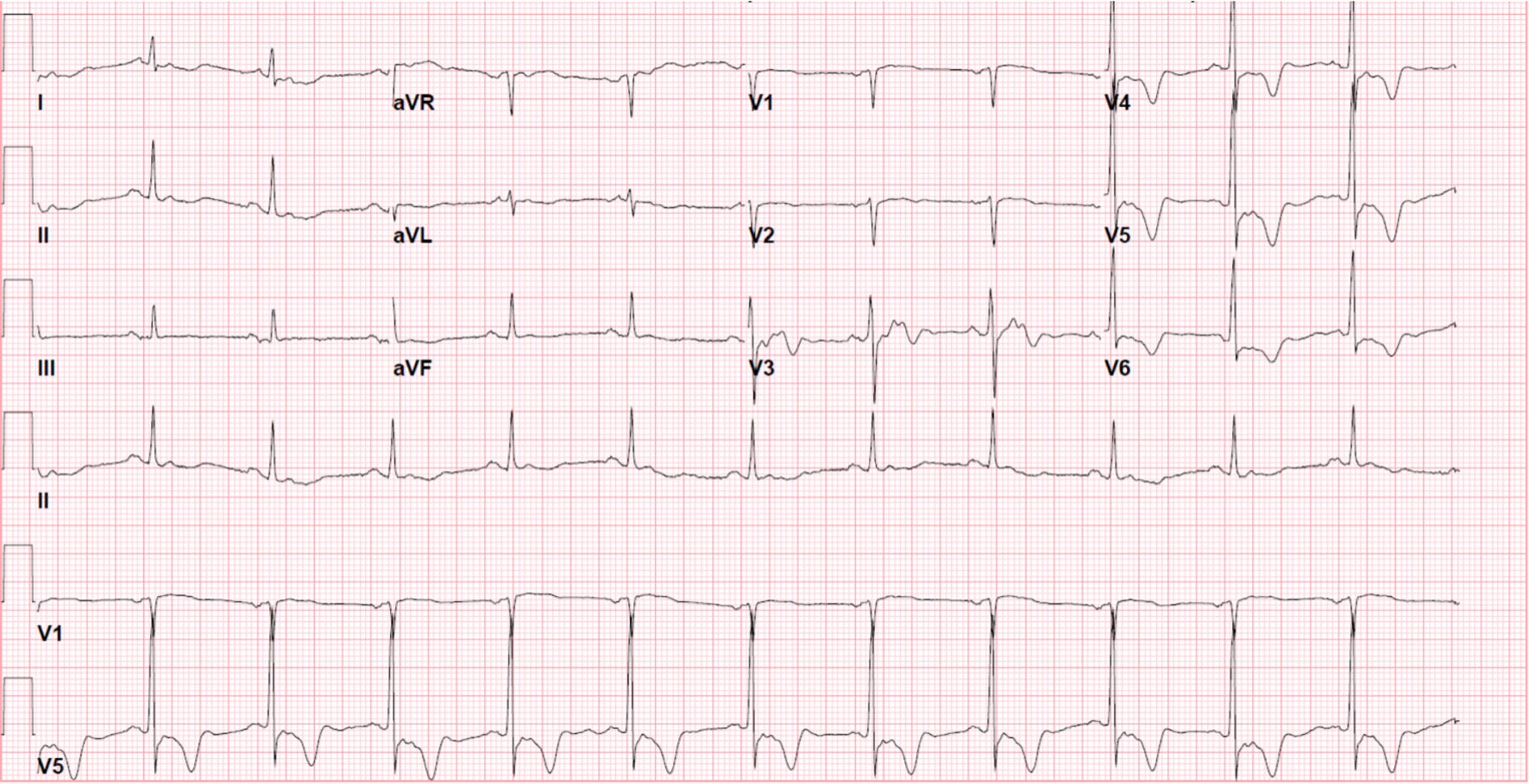
Figure 1. ECG on presentation shows atrial fibrillation and prominent T-waves inversion in leads V3 through V6. ECG: electrocardiogram.
| Cardiology Research, ISSN 1923-2829 print, 1923-2837 online, Open Access |
| Article copyright, the authors; Journal compilation copyright, Cardiol Res and Elmer Press Inc |
| Journal website https://www.cardiologyres.org |
Case Report
Volume 13, Number 6, December 2022, pages 393-397
Rare Occurrence of Apical Hypertrophic Cardiomyopathy Among Hispanics
Figures