Cardiology Research, ISSN 1923-2829 print, 1923-2837 online, Open Access
Article copyright, the authors; Journal compilation copyright, Cardiol Res and Elmer Press Inc
Journal website https://www.cardiologyres.org

Review

Volume 13, Number 4, August 2022, pages 177-184


Arrhythmogenic Right Ventricular Cardiomyopathy: The Role of Genetics in Diagnosis, Management, and Screening

Figures

Figure 1.
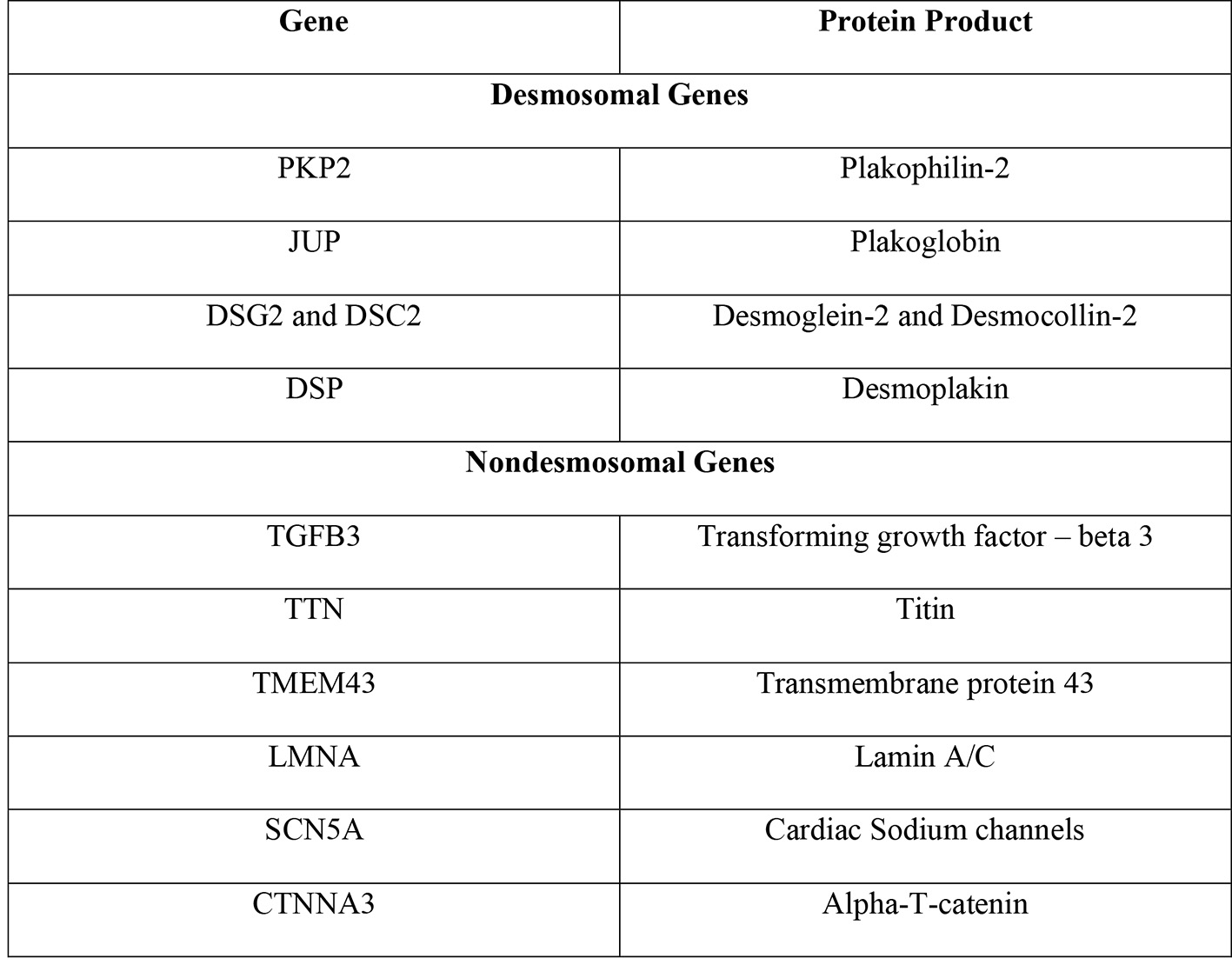
Figure 1. Genes implicated in ARVC and their protein products [8]. ARVC: arrhythmogenic right ventricular cardiomyopathy.
Figure 2.
Figure 2. The cardiac desmosome complex and the proteins involved [8].
Figure 3.
Figure 3. Revised Task Force Criteria for ARVC. *Corrected for body size (PLAX/BSA) ≥ 19 mm/m2 for major criteria and ≥ 16 to ≤ 18 mm/m2 for minor criteria. **Corrected for body size (PSAX/BSA) ≥ 21 mm/m2 for major criteria and ≥ 18 to < 21 mm/m2 for minor criteria [41]. ARVC: arrhythmogenic right ventricular cardiomyopathy; PLAX: parasternal long-axis view; RVOT: right ventricular outflow tract; RV: right ventricle; BSA: body surface area; PSAX: parasternal short-axis view; EDV: end-diastolic volume; EF: ejection fraction.
Figure 4.
Figure 4. Electrocardiographic findings associated with ARVC. Epsilon waves are characteristic of the condition, indicated by the red arrow. Negative T-waves in V1-V3 are major criteria while prolonged terminal activation duration of greater than 55 ms is a minor criterion for ARVC. ARVC: arrhythmogenic right ventricular cardiomyopathy.