Cardiology Research, ISSN 1923-2829 print, 1923-2837 online, Open Access
Article copyright, the authors; Journal compilation copyright, Cardiol Res and Elmer Press Inc
Journal website http://www.cardiologyres.org

Review

Volume 11, Number 4, August 2020, pages 205-212


Causes, Diagnosis, Risk Stratification and Treatment of Bicuspid Aortic Valve Disease: An Updated Review

Figure

Figure 1.
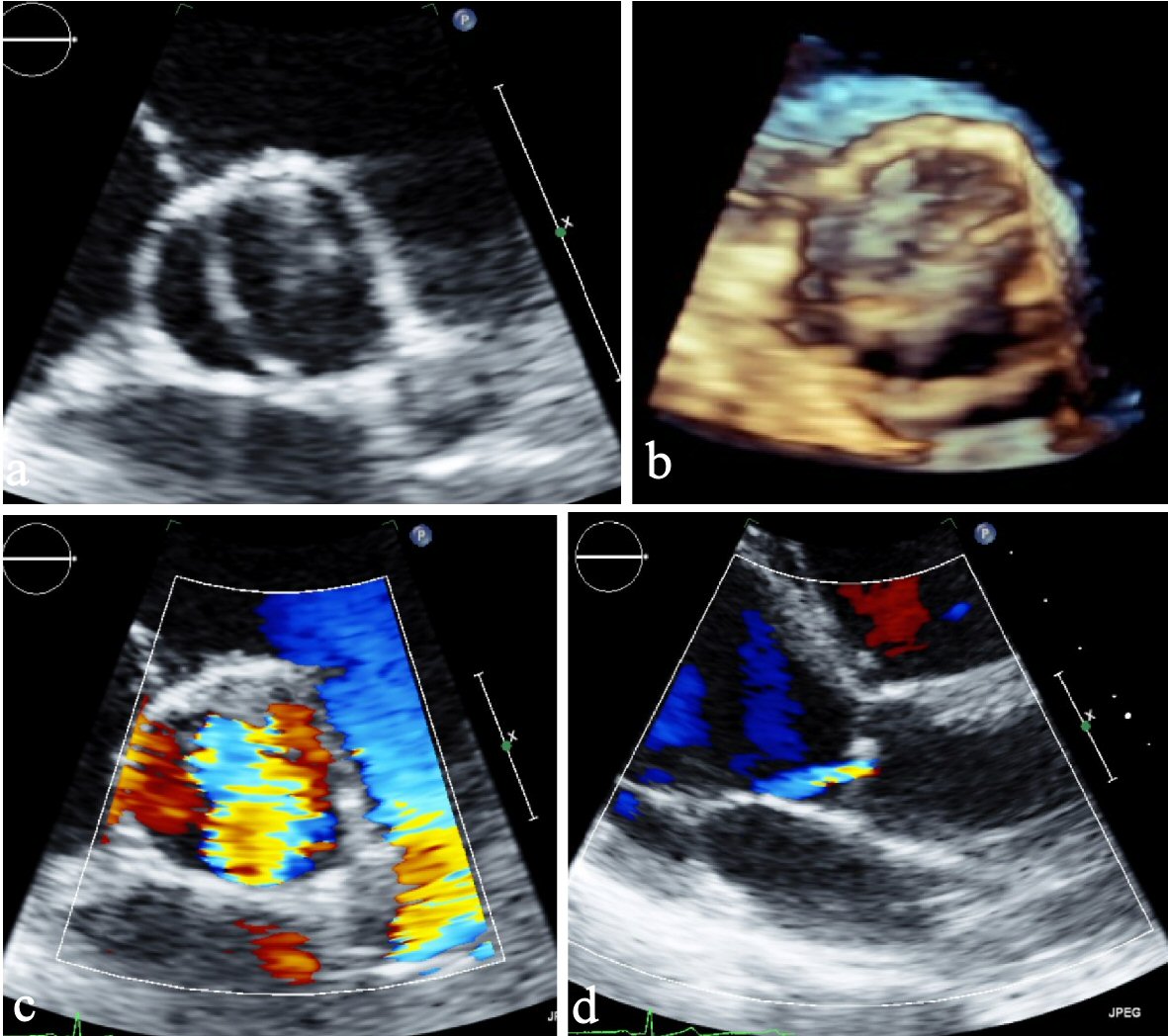
Figure 1. (a) Parasternal short axis view. (b) Parasternal short axis view (3D). (c) Parasternal short axis view of color Doppler showing severe aortic insufficiency. (d) Parasternal long axis view showing eccentric jet of regurgitant flow typical by BAV. BAV: bicuspid aortic valve; 3D: three-dimensional.

Table

Table 1. Screening and Follow-Up for Patients With BAV
 
TTE-screening of the aortic root and aorta ascendens for every patient with BAV should be performed. CT and MRI for precision diagnosis in case of inadequate TTE imaging.
If aortic root or aorta ascendens diameter is > 45 mm, or there is increase of 3 mm per year, follow-up every year is indicated.
In case of a diameter > 50 mm or there is increase of 3 mm per year in echocardiography, CT or MRI for confirmation should be performed.
Patients without significant heart valve lesions and aortic root diameter < 40 mm, cardiac imaging every 2 years may be sufficient.