Cardiology Research, ISSN 1923-2829 print, 1923-2837 online, Open Access
Article copyright, the authors; Journal compilation copyright, Cardiol Res and Elmer Press Inc
Journal website http://www.cardiologyres.org

Original Article

Volume 11, Number 3, June 2020, pages 179-184


Is the Time of Atrial Fibrillation Recurrence After Duty-Cycled Radiofrequency Ablation Affected by the Pattern of Pulmonary Vein Reconnections?

Figures

Figure 1.
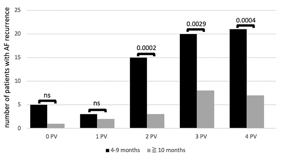
Figure 1. Number of patients with recurrence of AF during follow-up depending on number of reconnected PVs (0 - 4) found at redo PVI at different time intervals: early recurrence (black column) versus late recurrence (grey column). P values above each column. Left common trunk counted as one PV. AF: atrial fibrillation; ns: not significant: PV: pulmonary vein; PVI: pulmonary vein isolation.
Figure 2.
Figure 2. Correlation between time of AF recurrence after PVAC PVI and number of reconnected PVs at redo ablation with corresponding regression line. Left common trunk counted as one PV. PV: pulmonary vein; PVI: pulmonary vein isolation; AF: atrial fibrillation; PVAC: multielectrode duty-cycled radiofrequency.

Tables

Table 1. Clinical Characteristics of the Enrolled Patients (n = 85)
 
Data are numbers with mean ± SD. AF: atrial fibrillation; SD: standard deviation.
Age (years)58 ± 17 (31 - 72)
Male56 (66%)
Left atrial diameter (mm)43 ± 4 (37 - 50)
CHA2DS2-VASc-score2.2 (0 - 5)
Mean interval until first AF recurrence8.1 ± 3.4
Time interval until re-ablation (months)9.2 ± 3.8
Number of antiarrhythmic drugs1.4 ± 1.3 (0 - 3)
Amiodarone therapy17 (20%)
Hypertension42 (49%)
Coronary artery disease22 (26%)
No structural heart disease44 (52%)
Left ventricular ejection fraction (%)57 ± 8


Table 2. Electrophysiological Characteristics at Redo Procedure
 
PVAC (n = 85)
aIn patients with a reconnected common trunk, CT was considered and counted as one PV. Pts: patients; PVs: pulmonary veins; CTs: common trunks; LSPV: left superior pulmonary vein; LIPV: left inferior pulmonary vein; RSPV: right superior pulmonary vein; RIPV: right inferior pulmonary vein; PVAC: multielectrode duty-cycled radiofrequency.
Angiographically delineated PVs/CTs308/17
Targeted PVs/CTs at initial procedure302/17
Isolated PVs/CTs at initial procedure298/17
Pts. with PV reconnections79 (93%)
Reconnected PVs and CTs at redo procedure237/315 (75%)
  Reconnected CTs16/17 (94%)
  Reconnected LSPV57/67 (85%)
  Reconnected LIPV52/68 (76%)
  Reconnected RSPV63/79 (80%)
  Reconnected RIPV49/84 (58%)
PV reconnections per patienta2.79 ± 1.2
  Pts. with four reconnected PVs28 (33%)
  Pts. with three reconnected PVs28 (33%)
  Pts. with two reconnected PVs18 (21%)
  Pts. with one reconnected PV5 (6%)
  Pts. without reconnected PVs6 (7%)


Table 3. Time of AF Recurrence and Number of Reconnected PV at Redo Ablation in 85 Patients
 
4 - 6 months7 - 9 months10 - 12 months> 12 months
PV: pulmonary vein; AF: atrial fibrillation.
0 PV1401
1 PV1211
2 PVs31230
3 PVs81235
4 PVs15634