Figures
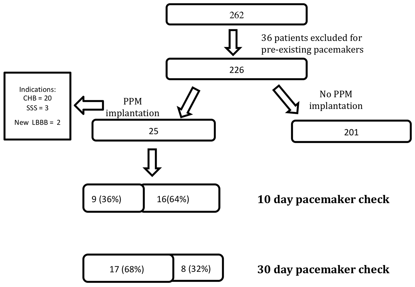
Figure 1. A total of 262 patients underwent transcatheter aortic valve replacement (TAVR), 36 patients were excluded from analysis because of pre-existing pacemakers and 25 patients required pacemaker implantation after TAVR for three different indications. All patients received a pacemaker check at 10 and 30 days. At 10 days, nine patients were no longer right ventricular (RV) pacing-dependent. At 30 days, 17 patients were no longer RV pacing-dependent. CHB: complete heart block; SSS: sick sinus syndrome; LBBB: left bundle branch block; PPM: permanent pacemaker.
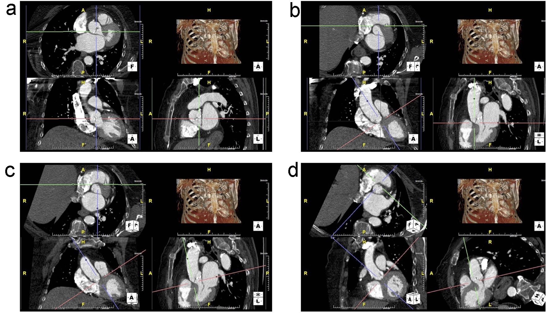
Figure 2. (a) Unaligned view of the aortic annulus that is not in plane. The top-left is the axial view, bottom-left is the transverse view and the bottom-right is the sagittal view. (b) The right coronary cusp (RCC) is now visible in the axial view by adjusting the red axes. The green and purple axes are then centered on the insertion of the RCC. (c) The non-coronary cusp (NCC) insertion is aligned by adjusting the red axes in the sagittal image to bring it into plane with the RCC insertion in the axial view. (d) The purple axes are adjusted to run through both RCC and NCC insertions. Then, the red axes on the transverse view are adjusted to bring in the insertion of the left coronary cusp (LCC) into the same plane as the RCC and NCC.
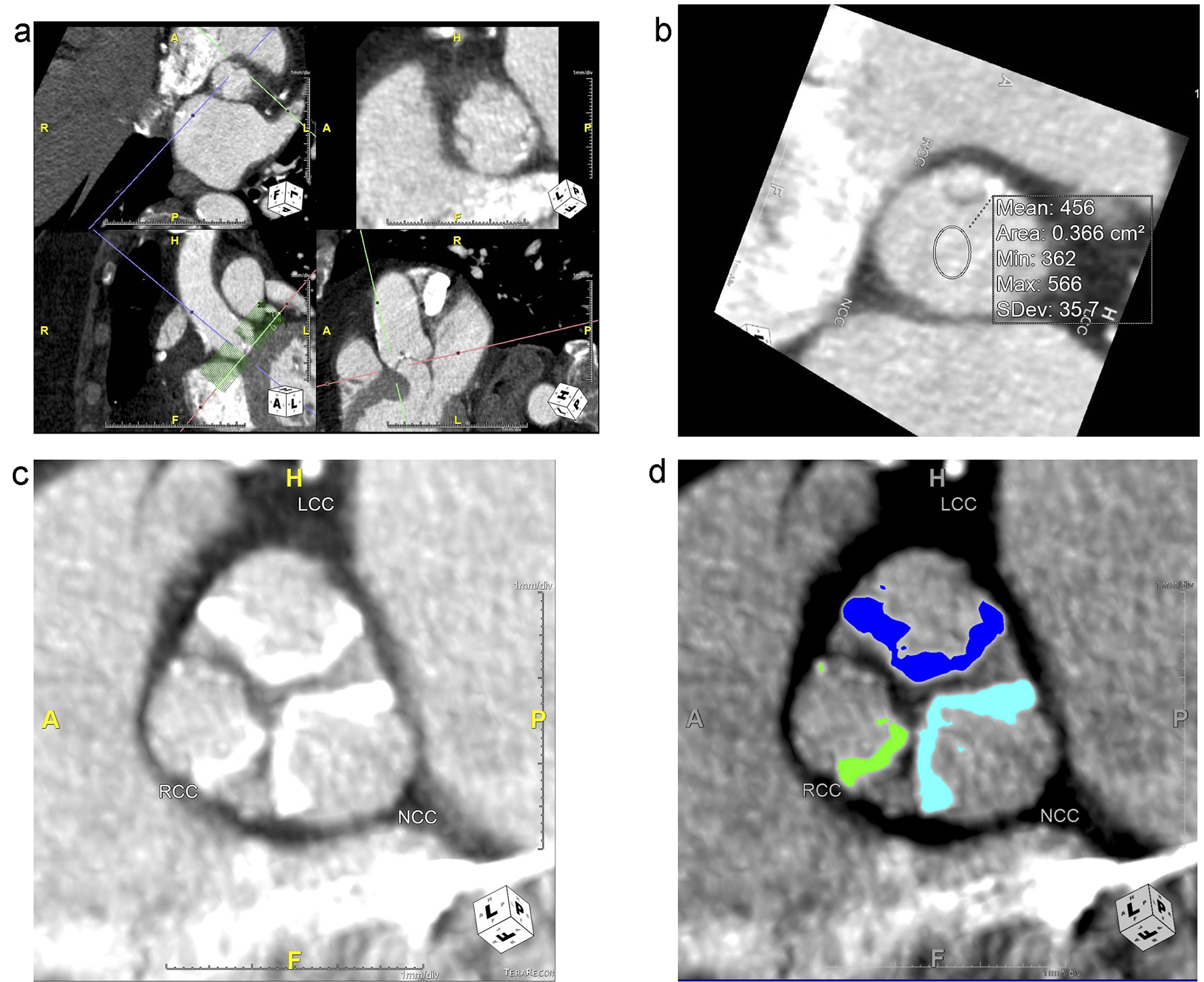
Figure 3. (a) The aortic valve complex used for analysis is created using a batching tool to separate the study images from the larger, original. The batching tool is visible in the bottom left in the axial view as a box of green lines, and it is centered at the annular plane (red axes). It extends 5 mm caudally from the annular plane and cranially to the tip of the upper leaflets. Each green line represents 1 mm. The output at the level of the annular plane is in the top-right box, with the cusps labeled as right coronary cusp (RCC), left coronary cusp (LCC) and non-coronary cusp (NCC) for orientation. (b) Tool used to calculate mean attenuation in Hounsfield Units (HU) to adjust cutoffs for calcium scoring. In this image, the mean luminal attenuation is 456 HU, so a cutoff of 550 HU was used. (c) Zoomed in view of leaflet calcification in one slice of the upper leaflet zone for each leaflet. (d) Calcification of each leaflet in this slice in the upper leaflet zone was traced using calcium analysis software, which then provided a calcium score, volume and mean. In the device landing zone and annulus region, a total calcium score, volume and mean was provided.
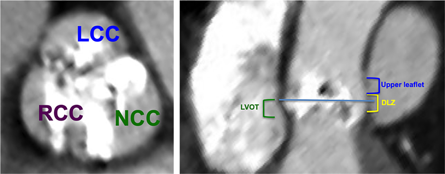
Figure 4. The aortic valve complex was separated in the craniocaudal axis of the left ventricular outflow tract (LVOT)/aortic valve into the following regions: the LVOT region (from 5 mm below the annular plane to the annular plane, green), the device-landing zone (2 mm inferior to the annular plane to 3 mm superior to the annular plane, yellow) and the upper leaflet region (3 mm superior to the annular plan to the superior edge of the leaflets, blue). Each leaflet was separately quantified in the upper leaflet region. NCC: non-coronary cusp; LCC: left coronary cusp; RCC: right coronary cusp.
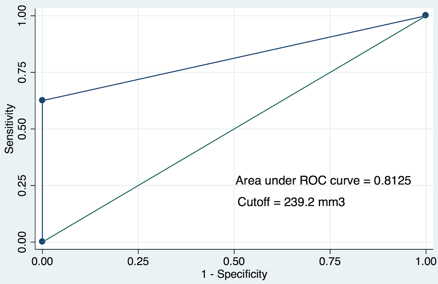
Figure 5. The receiver operating characteristic (ROC) curve for non-coronary cusp (NCC) volume and pacemaker dependence at 30 days. A cutoff of 239.2 mm3 for calcium was used.
Tables
Table 1. Electrocardiographic Data, Clinical Imaging Data and Procedural Parameters of Non-RV Pacing-Dependent and RV Pacing-Dependent Patients at 1 Month Post-PPM Implant
| Non-RV pacing-dependent (n = 17) | RV pacing-dependent (n = 8) | P-value |
|---|
| The P-values shown are derived from two-tailed Fisher’s exact test for categorical variables and two-tailed unpaired Student’s t-test for continuous variables. Patients who were non-RV pacing-dependent at 30 days and patients who were RV pacing-dependent at 30 days patients were separately compared with the non-pacemaker population to derive these P-values. Continuous variables are reported as the mean plus or minus the standard deviation. aValues obtained by transthoracic echocardiography. bValues obtained by multidetector computed tomography. RV: right ventricular; PPM: permanent pacemaker; AV: atrioventricular; LAFB: left anterior fascicular block; LPFB: left posterior fascicular block; RBBB: right bundle branch block; LBBB: left bundle branch block; LVEF: left ventricular ejection fraction; LVEDd: left ventricular end diastolic diameter; LVOTd: left ventricular outflow tract diameter; MAC: mitral annular calcification; TAVR: transcatheter aortic valve replacement; CHB: complete heart block; SSS: sick sinus syndrome; LVOT: left ventricular outflow tract. |
| Electrocardiographic data | | | |
| Sinus | 11/17 | 7/8 | 0.36 |
| First-degree AV block | 3/17 | 2/8 | 1 |
| LAFB | 4/17 | 3/8 | 0.64 |
| LPFB | 1/17 | 1/8 | 1 |
| RBBB | 2/17 | 8/8 | < 0.0001 |
| LBBB | 1/17 | 0/8 | 1 |
| Bifascicular block | 2/17 | 5/8 | 0.017 |
| QRS duration > 120 ms | 3/17 | 8/8 | 0.0002 |
| QRS duration (ms) | 108.4 ± 22.0 | 138.8 ± 14.1 | 0.0017 |
| Clinical imaging data | | | |
| Aortic valve areaa | 0.65 ± 0.23 | 0.77 ± 0.26 | 0.23 |
| LVEFa | 52.1 ± 13.9 | 59.4 ± 11.8 | 0.21 |
| LVOTda | 2.04 ± 0.33 | 2.10 ± 0.22 | 0.64 |
| LVEDda | 4.94 ± 0.80 | 4.64 ± 0.84 | 0.43 |
| Aortic annulus diameterb | 2.37 ± 0.30 | 2.51 ± 0.22 | 0.25 |
| Aortic annulus areab | 4.48 ± 1.12 | 5.06 ± 0.79 | 0.21 |
| Severe MAC | 4/17 | 0/8 | 0.28 |
| Procedural parameters | | | |
| Transfemoral approach | 13/17 | 8/8 | 0.27 |
| Transapical approach | 4/17 | 0/8 | 0.27 |
| Intra-procedural complete heart block | 6/17 | 7/8 | 0.03 |
| Valve size | 25.3 ± 2.5 | 25.3 ± 2.1 | 0.97 |
| Valve/LVOTda | 12.6 ± 1.8 | 12.1 ± 1.1 | 0.45 |
| Days post-TAVR of pacemaker insertion | 5.24 ± 3.44 | 5.88 ± 6.31 | 0.74 |
| Paravalvular leak requiring post-dilation | 7/17 | 1/8 | 0.21 |
| Percent oversizing by annulus areab | 13.7 ± 15.3 | -0.03 ± 12.7 | 0.04 |
| Generation | | | |
| SAPIEN | 12/17 | 5/8 | 1 |
| SAPIEN XT | 3/17 | 0/8 | 0.53 |
| SAPIEN III | 2/17 | 3/8 | 0.28 |
| Pacemaker indication | | | |
| CHB | 12/17 | 8/8 | 0.14 |
| SSS | 3/17 | 0/8 | 0.53 |
| New LBBB | 2/17 | 0/8 | 0.55 |
Table 2. Baseline Characteristics and Comparison of Patients Not Requiring PPM Implant and Those Requiring PPM Implant Post-TAVR
| No PPM (n = 201) | Non-RV pacing-dependent at 30 days (n = 17) | P-value | Pacing-dependent at 30 days (n = 8) | P-value |
|---|
| The P-values shown are derived from two-tailed Fisher’s exact test for categorical variables and two-tailed unpaired Student’s t-test for continuous variables. Patients who were non-RV pacing-dependent at 30 days and patients who were RV pacing-dependent at 30 days patients were separately compared with the non-pacemaker population to derive these P-values. Continuous variables are reported as the mean plus or minus the standard deviation. PPM: permanent pacemaker; TAVR: transcatheter aortic valve replacement; RV: right ventricular; BMI: body mass index; DM: diabetes mellitus; CAD: coronary artery disease; CHF: congestive heart failure; COPD: chronic obstructive pulmonary disease; PVD: peripheral vascular disease; eGFR: estimated glomerular filtration rate; CABG: coronary artery bypass grafting; PCI: percutaneous coronary intervention; ACE-I/ARB: angiotensin converting enzyme inhibitor/angiotensin receptor blocker; AV: atrioventricular; LAFB: left anterior fascicular block; LPFB: left posterior fascicular block; RBBB: right bundle branch block; LBBB: left bundle branch block; LVEF: left ventricular ejection fraction; LVOTd: left ventricular outflow tract diameter. |
| Age | 82.2 ± 8.3 | 80.4 ± 6.3 | 0.39 | 80.9 ± 8.3 | 0.66 |
| Gender (female) | 102/201 | 8/17 | 0.81 | 2/8 | 0.28 |
| BMI | 27.2 ± 6.3 | 29.1 ± 6.3 | 0.27 | 25.0 ± 4.5 | 0.32 |
| Hypertension | 185/201 | 15/17 | 0.64 | 5/8 | 0.03 |
| DM | 70/201 | 10/17 | 0.066 | 2/8 | 0.72 |
| CAD | 157/201 | 14/17 | 0.77 | 5/8 | 0.38 |
| CHF | 142/201 | 13/17 | 0.78 | 5/8 | 0.70 |
| COPD | 37/201 | 2/17 | 0.55 | 1/8 | 1 |
| PVD | 29/201 | 4/17 | 0.48 | 1/8 | 1 |
| History of atrial fibrillation | 69/201 | 6/17 | 1 | 2/8 | 0.72 |
| History of atrial flutter | 26/175 | 1/17 | 0.49 | 1/8 | 1 |
| eGFR < 60 | 97/201 | 10/17 | 0.46 | 7/8 | 0.035 |
| Creatinine | 1.12 ± 0.54 | 1.36 ± 0.61 | 0.09 | 1.92 ± 1.42 | 0.0002 |
| Prior CABG | 66/201 | 7/17 | 0.59 | 2/8 | 0.73 |
| Prior PCI | 74/201 | 3/17 | 0.12 | 3/8 | 1 |
| Prior balloon valvuloplasty | 25/201 | 5/17 | 0.065 | 0/8 | 0.60 |
| ACE-I/ARB | 106/201 | 7/17 | 0.45 | 5/8 | 0.73 |
| Beta-blockers | 144/201 | 10/17 | 0.28 | 6/8 | 1 |
| Digoxin | 13/201 | 0/17 | 0.41 | 0/8 | 1 |
| Other anti-arrhythmic drugs | 12/201 | 0/17 | 0.61 | 0/8 | 1 |
| Sinus rhythm | 161/200 | 11/17 | 0.21 | 7/8 | 1 |
| First-degree AV block | 42/200 | 3/17 | 1 | 2/8 | 1 |
| LAFB | 27/200 | 4/17 | 0.28 | 3/8 | 0.09 |
| LPFB | 1/200 | 1/17 | 0.15 | 1/8 | 0.08 |
| RBBB | 18/200 | 2/17 | 1 | 8/8 | < 0.0001 |
| LBBB | 22/200 | 1/17 | 0.70 | 0/8 | 0.60 |
| Bifascicular block | 9/200 | 2/17 | 0.21 | 5/8 | < 0.0001 |
| QRS duration > 120 ms | 48/200 | 3/17 | 0.77 | 8/8 | < 0.0001 |
| QRS duration (ms) | 106.1 ± 24.2 | 108.4 ± 22.0 | 0.71 | 138.8 ± 14.1 | 0.0002 |
| Aortic valve area | 0.66 ± 0.23 | 0.65 ± 0.23 | 0.78 | 0.77 ± 0.26 | 0.19 |
| LVEF | 56.4 ± 13.4 | 52.1 ± 13.9 | 0.21 | 59.4 ± 11.8 | 0.53 |
| LVOTd | 2.02 ± 0.22 | 2.04 ± 0.33 | 0.74 | 2.1 ± 0.22 | 0.31 |
| Transfemoral approach | 155/201 | 13/17 | 1 | 8/8 | 0.20 |
| Transapical approach | 36/201 | 4/17 | 0.74 | 0/8 | 0.36 |
| Intra-procedural complete heart block | 17/201 | 6/17 | 0.004 | 7/8 | < 0.0001 |
| Valve size | 25.0 ± 2.3 | 25.3 ± 2.5 | 0.60 | 25.3 ± 2.1 | 0.75 |
| Valve/LVOTd | 12.5 ± 1.3 | 12.6 ± 1.8 | 0.71 | 12.1 ± 1.1 | 0.38 |
| Percent oversizing by annulus area | 11.3 ± 18.0 | 13.7 ± 15.3 | 0.60 | -0.03 ± 12.7 | 0.08 |
Table 3. Baseline Demographic and Clinical Characteristics of Non-RV Pacing-Dependent and Pacing-Dependent Patients at 1 Month Post-PPM Implant
| Non-RV pacing-dependent (n = 17) | RV pacing-dependent (n = 8) | P-value |
|---|
| The P-values shown are derived from two-tailed Fisher’s exact test for categorical variables and two-tailed unpaired Student’s t-test for continuous variables. Patients who were non-RV pacing-dependent at 30 days and patients who were RV pacing-dependent at 30 days patients were separately compared with the non-pacemaker population to derive these P-values. Continuous variables are reported as the mean plus or minus the standard deviation. RV: right ventricular; PPM: permanent pacemaker; BMI: body mass index; DM: diabetes mellitus; CAD: coronary artery disease; CHF: congestive heart failure; COPD: chronic obstructive pulmonary disease; PVD: peripheral vascular disease; eGFR: estimated glomerular filtration rate; CABG: coronary artery bypass grafting; PCI: percutaneous coronary intervention; ACE-I/ARB: angiotensin converting enzyme inhibitor/angiotensin receptor blocker. |
| Age | 80.4 ± 6.3 | 80.9 ± 8.3 | 0.88 |
| Gender (female) | 8/17 | 2/8 | 0.40 |
| BMI | 29.1 ± 6.3 | 25.0 ± 4 .5 | 0.12 |
| Hypertension | 15/17 | 5/8 | 0.28 |
| DM | 10/17 | 2/8 | 0.2 |
| CAD | 14/17 | 5/8 | 0.34 |
| CHF | 13/17 | 5/8 | 0.64 |
| COPD | 2/17 | 1/8 | 1 |
| PVD | 4/17 | 1/8 | 0.64 |
| History of atrial fibrillation | 6/17 | 2/8 | 0.68 |
| History of atrial flutter | 1/17 | 1/8 | 1 |
| eGFR < 60 | 10/17 | 7/8 | 0.21 |
| Creatinine | 1.36 ± 0.61 | 1.92 ± 1.42 | 0.19 |
| Prior CABG | 7/17 | 2/8 | 0.66 |
| Prior PCI | 3/17 | 3/8 | 0.34 |
| Prior balloon valvuloplasty | 5/17 | 0/8 | 0.14 |
| ACE-I/ARB | 7/17 | 5/8 | 0.41 |
| Beta-blockers | 10/17 | 6/8 | 0.66 |
| Digoxin | 0/17 | 0/8 | 1 |
| Other anti-arrhythmic drugs | 0/17 | 0/8 | 1 |
Table 4. Aortic Valve Complex Calcification
| RV pacing-dependent at 30 days (n = 8) | Non-RV pacing-dependent at 30 days (n = 16) | P-value |
|---|
| The P-values shown are derived from two-tailed unpaired Student’s t-test for continuous variables. Patients who were non-RV pacing-dependent at 30 days and patients who were RV pacing-dependent at 30 days patients were separately compared. Continuous variables are reported as the mean plus or minus the standard deviation. HU: Hounsfield Units; LVOT: left ventricular outflow tract; RCC: right coronary cusp; NCC: non-coronary cusp; LCC: left coronary cusp; RV: right ventricular. |
| Mean attenuation | 336.6 ± 87.6 | 353.8 ± 120.2 | 0.72 |
| HU cutoff | 353.8 ± 70.7 | 539.8 ± 66.9 | 0.62 |
| Total calcium volume | 783.5 ± 622.1 | 420.3 ± 264.2 | 0.055 |
| LVOT calcium volume | 673.7 ± 202.8 | 697.6 ± 34.7 | 0.18 |
| Annulus calcium volume | 117.9 ± 120.6 | 92.9 ± 50.4 | 0.48 |
| RCC leaflet calcium volume | 119.7 ± 121.5 | 107.2 ± 119.9 | 0.81 |
| NCC leaflet calcium volume | 342.0 ± 290.8 | 113.6 ± 63.2 | 0.006 |
| LCC leaflet calcium volume | 139.9 ± 132.7 | 91.4 ± 87.0 | 0.29 |
| Total leaflet calcium volume | 601.6 ± 504.9 | 312.2 ± 248.8 | 0.07 |
Table 5. Predictors of Persistent RV Pacing After PPM Implantation
| RV pacing-dependent (n = 8) | Non-RV pacing-dependent (n = 17) | Odds ratio (95% CI) | P-value |
|---|
| The P-values shown are derived from two-tailed Fisher’s exact test for categorical variables. Patients who were non-RV pacing-dependent at 30 days and patients who were RV pacing-dependent at 30 days patients were separately compared. RBBB: right bundle branch block; CI: confidence interval; NCC: non-coronary cusp; RV: right ventricular; PPM: permanent pacemaker. |
| RBBB | 8/8 | 2/17 | 105.4 (4.52 - 2,458.5) | 0.004 |
| Bifascicular block | 5/8 | 2/17 | 12.50 (1.6 - 97.65) | 0.02 |
| QRS duration > 120 ms | 8/8 | 3/17 | 70.43 (3.23 - 1,535.22) | 0.007 |
| Intra-procedural complete heart block | 7/8 | 6/17 | 12.83 (1.26 - 130.52) | 0.03 |
| NCC volume > 239.2 mm3 | 6/8 | 0/16 | 85.80 (3.61 - 2,041.00) | 0.006 |