Cardiology Research, ISSN 1923-2829 print, 1923-2837 online, Open Access
Article copyright, the authors; Journal compilation copyright, Cardiol Res and Elmer Press Inc
Journal website https://www.cardiologyres.org

Case Report

Volume 9, Number 2, April 2018, pages 133-135


Mitroflow Aortic Bioprosthesis Failure in Type B Aortic Dissection: Preventive Left Main Stenting in Transapical Transcatheter Aortic Valve Implantation Strategy

Figures

Figure 1.
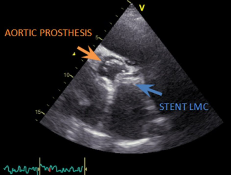
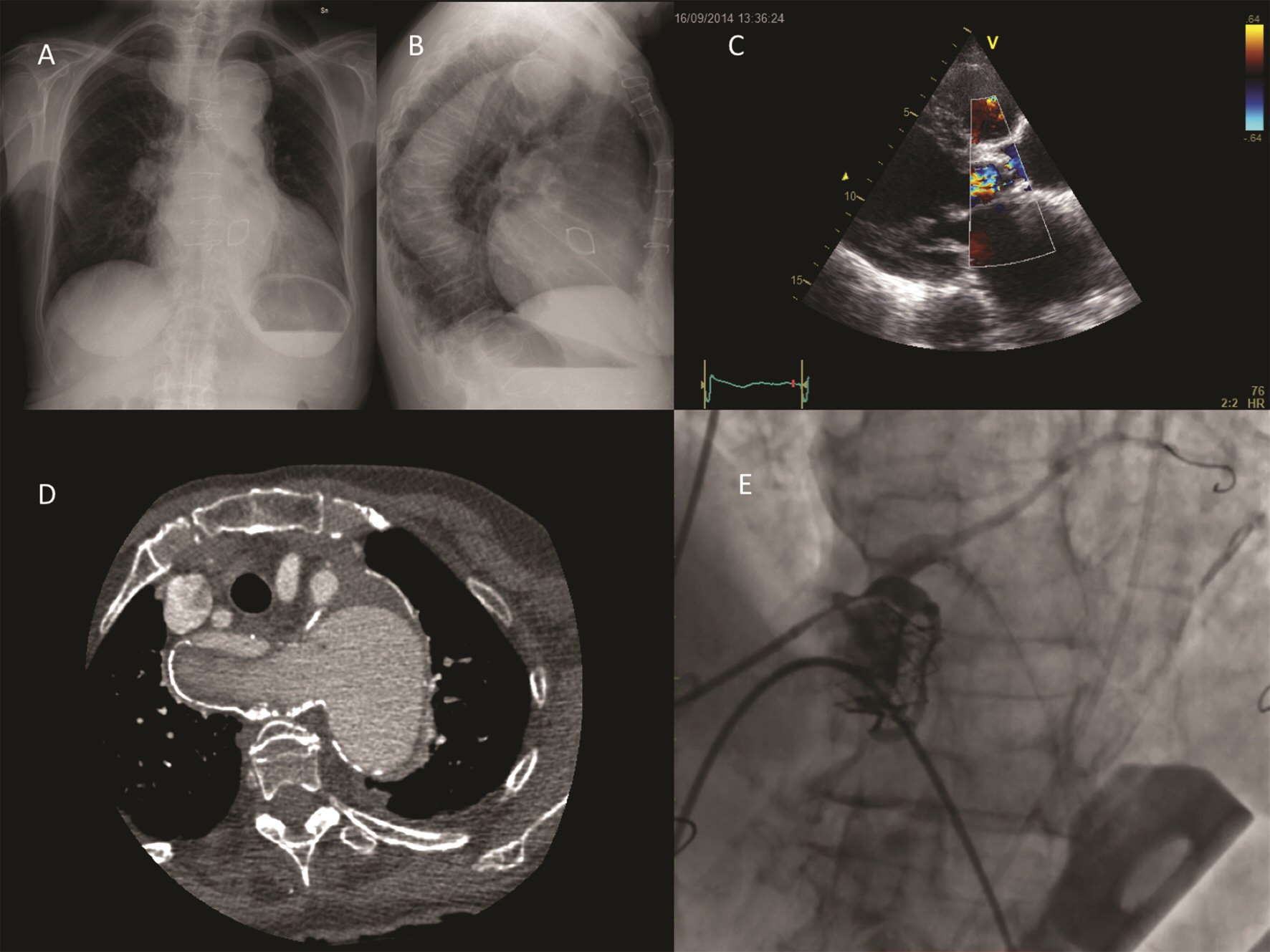
Figure 1. (A-B): Chest X-ray in posteroanterior and laterolateral view. Calcifications of the vascular knobs (aortic arch on the left, right subclavian artery from the diverticulum on the right) and of the descending aorta are evident. (C): Severe aortic regurgitation from Mitroflow prosthesis dysfunction. (D): CT scan shows chronic type B aortic dissection and the calcified Kommerell diverticulum. (E): Intraprocedural angiography shows stenting of the left main after transapical Sapien XT released.
Figure 2.
Figure 2. Late follow-up echocardiogram showing the correct positioning both of the TAVI prosthesis and of the stent implanted in the left main.