Cardiology Research, ISSN 1923-2829 print, 1923-2837 online, Open Access
Article copyright, the authors; Journal compilation copyright, Cardiol Res and Elmer Press Inc
Journal website https://www.cardiologyres.org

Original Article

Volume 8, Number 6, December 2017, pages 286-292


Transcatheter Closure Versus Repeat Surgery for the Treatment of Postoperative Left-to-Right Shunts: A Single Center 15-Year Experience

Figures

Figure 1.
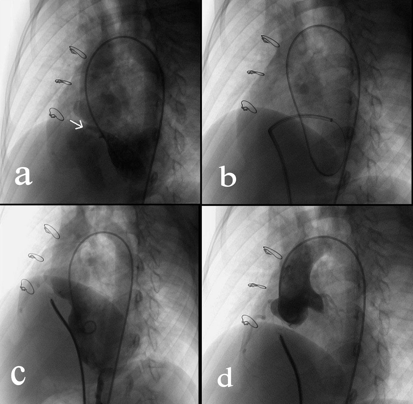
Figure 1. Transcatheter closure of a residual VSD. (a) Angiography of left ventricle via a 4F pigtail catheter inserted through the right femoral artery showed the residual VSD (indicated by the arrow). (b) Via the right femoral vein, a femoral vein-inferior vena cava-right ventricle-residual VSD-left ventricle-aorta-right femoral artery loop was established using a 260 cm loach guide wire. Afterwards, a 6-F delivery catheter was advanced along the loop to the left ventricle. (c) The loop wire was withdrawn. Subsequently, a VSD occluder was delivered to left ventricle and then opened in following sequence: left disc, waist and right disc. (d) After repeated angiography showed that the shunt disappeared, and then the occluder was released.
Figure 2.
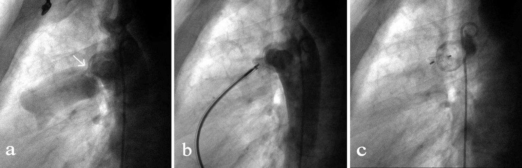
Figure 2. Transcatheter closure of a residual PDA. (a) Angiography of the aortic arch via a 5-F pigtail catheter that was inserted through the right femoral artery showed the residual PDA shunt (indicated by the arrow). (b) By the access of right femoral vein, a MPA2 catheter was advanced via right ventricle, pulmonary artery, PDA to the descending aorta followed by the insert of a 260 mm wire. After the MPA2 catheter was withdrawn, an 8-F delivery sheath was advanced along the wire and the duct occluder was delivered, positioned and opened in such a sequence that the aortic end of the occluder was first opened and then the pulmonary artery end. (c) Repeated angiography showed the occluder was properly placed and the shunt disappeared, and then the duct occluder was released.
Figure 3.
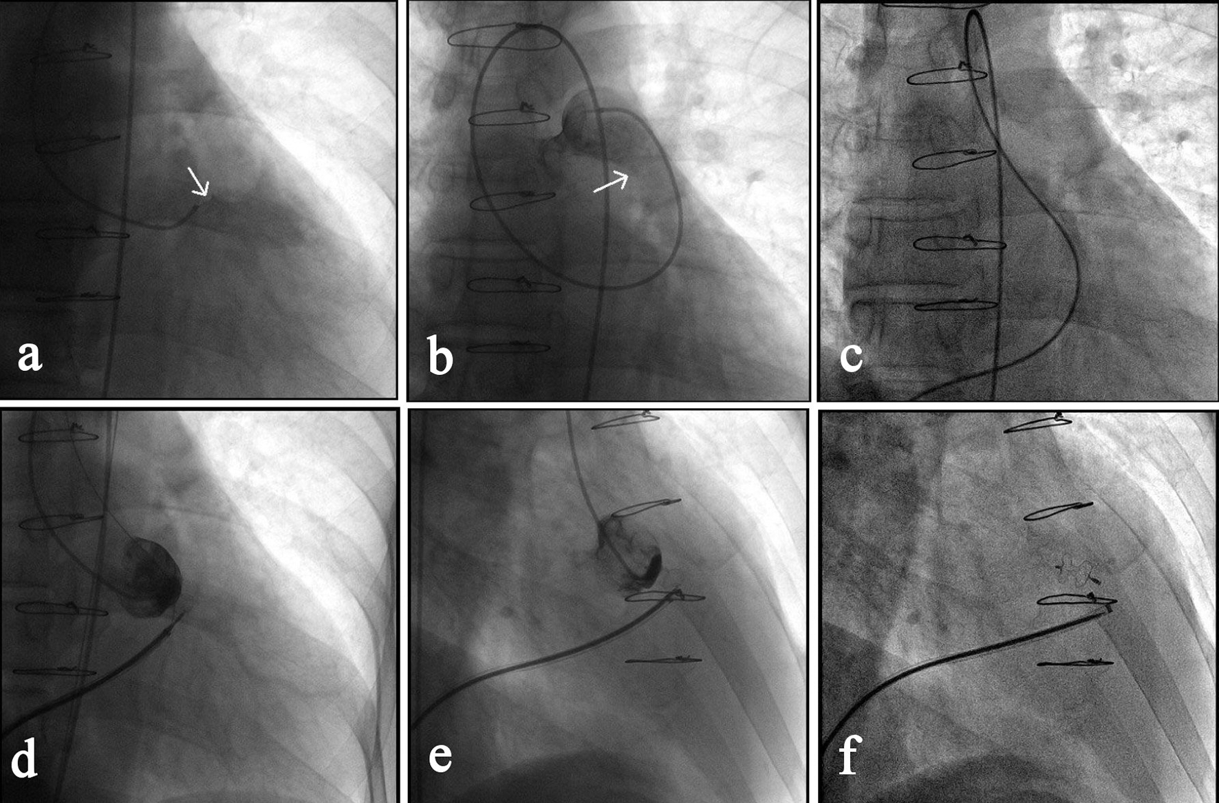
Figure 3. Transcatheter closure of a RSVA. (a) Angiography of right coronary sinus via a MPA1 catheter that was inserted into the right femoral artery showed the shunt from right coronary sinus to the right ventricular outflow tract (as indicated by the arrow). (b) Angiography after the MPA1 catheter was further advanced to the pulmonary artery confirmed the shunt (arrow). (c) Via the right femoral vein, a femoral vein-inferior vena cava-right ventricle-RSVA-aorta-right femoral artery loop was established using a 260 cm loach guide wire. (d) A 7-F delivery sheath was advanced along the loop to ascending aorta, and then a 7 mm VSD occluder was delivered passing the RSVA and opened in following sequence: left disc, waist and right disc. (e) The loop wire was withdrawn and angiography showed the shunt disappeared. (f) The occluder was released, delivery catheter withdrawn and repeated angiography showed the occluder was properly placed.

Tables

Table 1. Demographics and Clinical Characteristics of Patients Undergoing Transcatheter Closure
 
PatientAge (years)SexPrimary diagnosisShunt size (mm)Qp/QsPAP (mm Hg)Device type size (mm)Procedure time (min)Hospital stay (days)
VSD: ventricular septal defect; TOF: tetralogy of Fallot; PDA: patent ductus arteriosus; PAP: pulmonary arterial pressure.
17FVSD41.532/14 (22)pmVSD, 6566
226MVSD81.927/11 (16)pmVSD, 10627
314MVSD51.630/13 (19)pmVSD, 8457
429FVSD61.521/9 (12)pmVSD, 8916
530FVSD31.519/8 (11)pmVSD, 5676
63FVSD42.122/11 (15)pmVSD, 6467
728MVSD52.216/7 (10)pmVSD, 81127
812FVSD82.915/5 (9)pmVSD, 10576
98FVSD31.532/15 (23)pmVSD, 5366
1013MVSD41.738/17 (26)pmVSD, 7526
1159FVSD72.224/10 (21)pmVSD, 10717
124FVSD31.722/9 (14)pmVSD, 5457
139FVSD61.918/7 (12)pmVSD, 8396
1418MVSD122.817/8 (12)pmVSD, 141526
157FVSD41.814/6 (8)pmVSD, 6416
1638MTOF103.129/15 (21)pmVSD, 12976
1714MTOF41.635/18 (26)pmVSD, 6537
1845MVSD41.541/22 (30)pmVSD, 71086
195MVSD51.927/14 (19)PDA, 10/8416
2018FPDA31.526/12 (17)PDA, 6/4295
2121FPDA51.739/18 (28)PDA, 10/8395
2219FPDA41.831/15 (22)PDA, 8/6315
237MVSD41.726/13 (19)pmVSD, 7415
2446MPDA51.941/23 (31)PDA, 12/10396
2514FTOF41.630/13 (19)pmVSD, 8485
2665MVSD51.743/24 (32)mVSD, 12678
276FTOF31.621/9 (13)pmVSD, 5415
2821FTOF71.926/14 (17)pmVSD, 10526
299MVSD31.732/13 (22)pmVSD, 6375
3057MVSD61.849/27 (36)mVSD, 14786
318MVSD51.817/8 (12)pmVSD, 7526


Table 2. Demographics and Clinical Characteristics of Patients Treated With Reoperation
 
PatientAge (years)SexPrimary diagnosisShunt size (mm)PAP (mm Hg)ComplicationsProcedure time (min)Hospital stay (days)
VSD: ventricular septal defect; TOF: tetralogy of Fallot; DORV: double outlet right ventricle; B: bleeding; WD: wound dehiscence; P: pneumonia; KI: kidney insufficiency.
112FVSD521B21315
231MVSD427B28717
337FVSD621B, WD, P31421
44MVSD43118714
526FVSD719B26116
615MVSD921B, P34219
710FVSD32919813
89FVSD43123615
945MVSD641B, P32712
107FVSD41617817
1113FVSD617B22611
1221MVSD821B, KI29115
136FVSD32617010
1436FVSD1046B23513
159FTOF516B21714
1611MTOF51324116
176FTOF63133917
185MDORV426B, WD35828
1914FTOF71826118


Table 3. Summarized Data for Patients Undergoing TCC and Reoperation
 
Patients with TCCPatients with reoperation
*P < 0.001 compared with TCC group. N/A: not available. TTC: transcatheter closure.
Total number (before 2010)114
Total number (after 2010)305
SexFemale: 16; male: 15Female: 12; male: 7
Age (years)21.3 ± 17.316.7 ± 12.4
Average shunt size (mm)5.1 ± 2.15.6 ± 2.0
Qp/Qs1.9 ± 0.4N/A
Procedure time (min)58.9 ± 27.7256.7 ± 60.5*
Hospital stay (days)6.1 ± 0.817.0 ± 4.0*