Cardiology Research, ISSN 1923-2829 print, 1923-2837 online, Open Access
Article copyright, the authors; Journal compilation copyright, Cardiol Res and Elmer Press Inc
Journal website https://www.cardiologyres.org

Review

Volume 8, Number 4, August 2017, pages 139-142


Feline Hypertrophic Cardiomyopathy: A Spontaneous Large Animal Model of Human HCM

Figures

Figure 1.
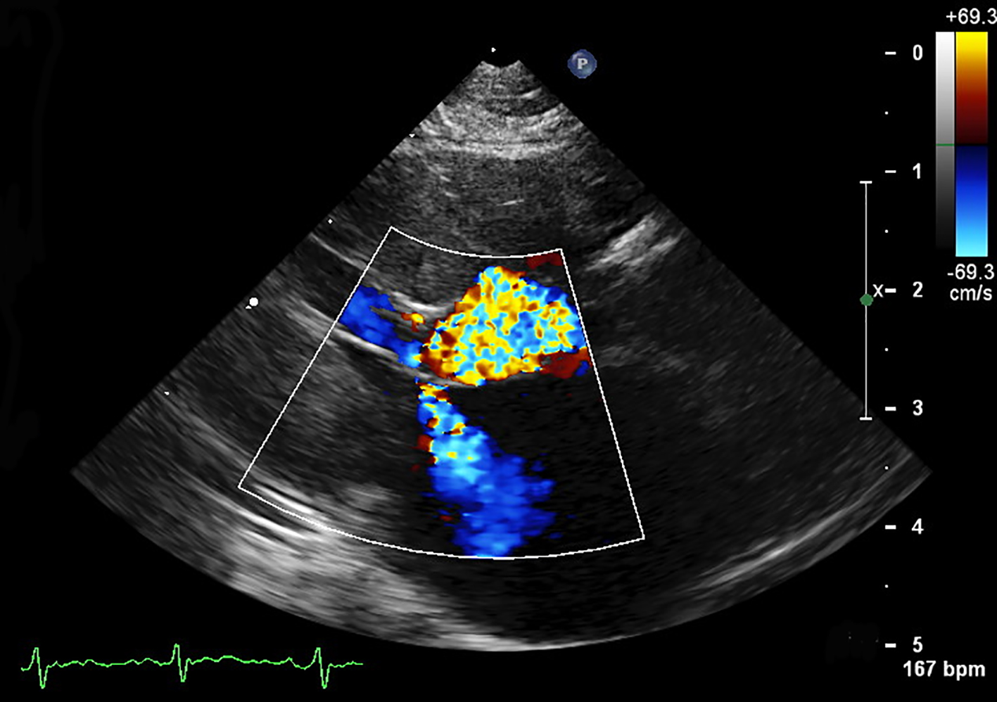
Figure 1. A right parasternal echocardiogram with color flow Doppler from a 7-year-old cat with hypertrophic obstructive cardiomyopathy. Mitral regurgitation and left ventricular outflow tract obstruction secondary to systolic anterior motion of the mitral valve are seen. This patient has significant left ventricular hypertrophy of the interventricular septum and free wall.
Figure 2.
Figure 2. A gross pathology specimen from a 5-year-old cat with hypertrophic cardiomyopathy. The heart is shown in a transverse plane at the level of the papillary muscles in the left ventricle. Severe concentric left ventricular hypertrophy is evident with dramatic reduction in the size of the left ventricular lumen.