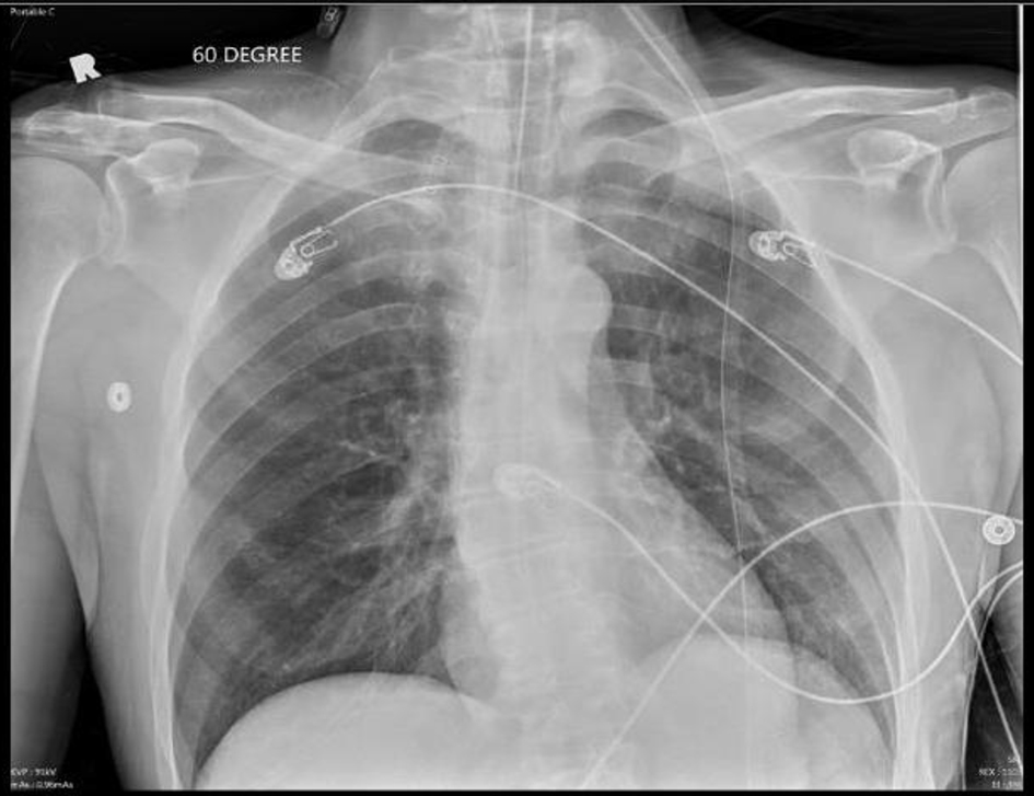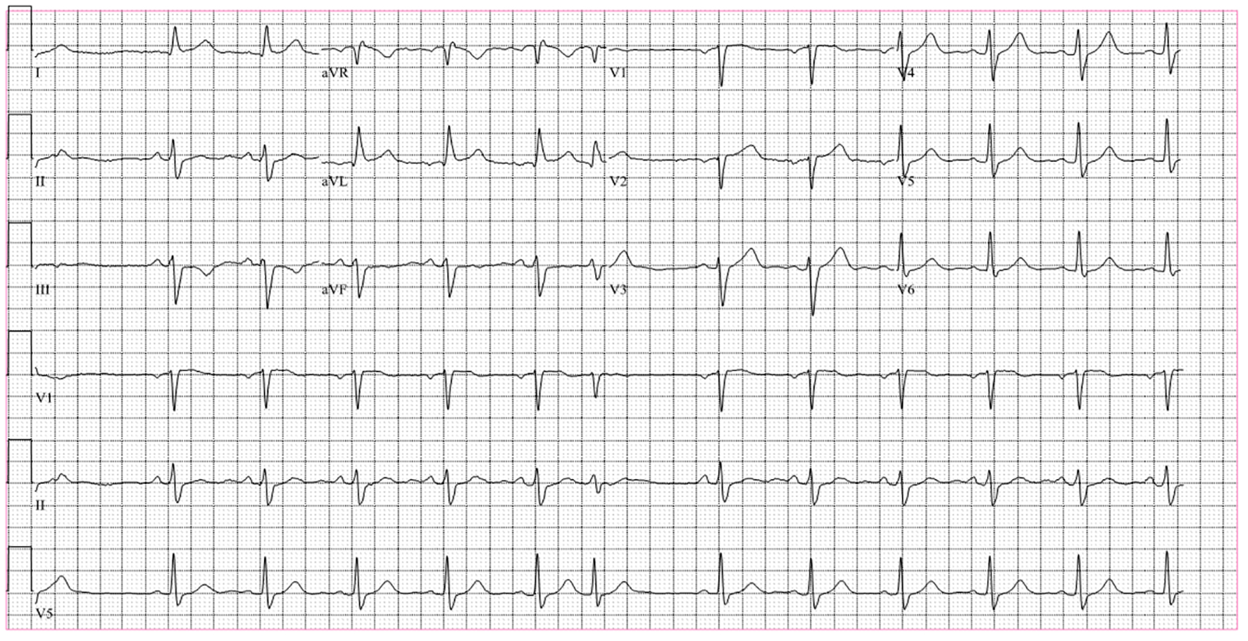
Figure 1. Baseline EKG on admission shows normal sinus rhythm with few premature ventricular contractions. QT interval is 432 ms and corrected QT is 464 ms.
| Cardiology Research, ISSN 1923-2829 print, 1923-2837 online, Open Access |
| Article copyright, the authors; Journal compilation copyright, Cardiol Res and Elmer Press Inc |
| Journal website https://www.cardiologyres.org |
Case Report
Volume 8, Number 5, October 2017, pages 232-235
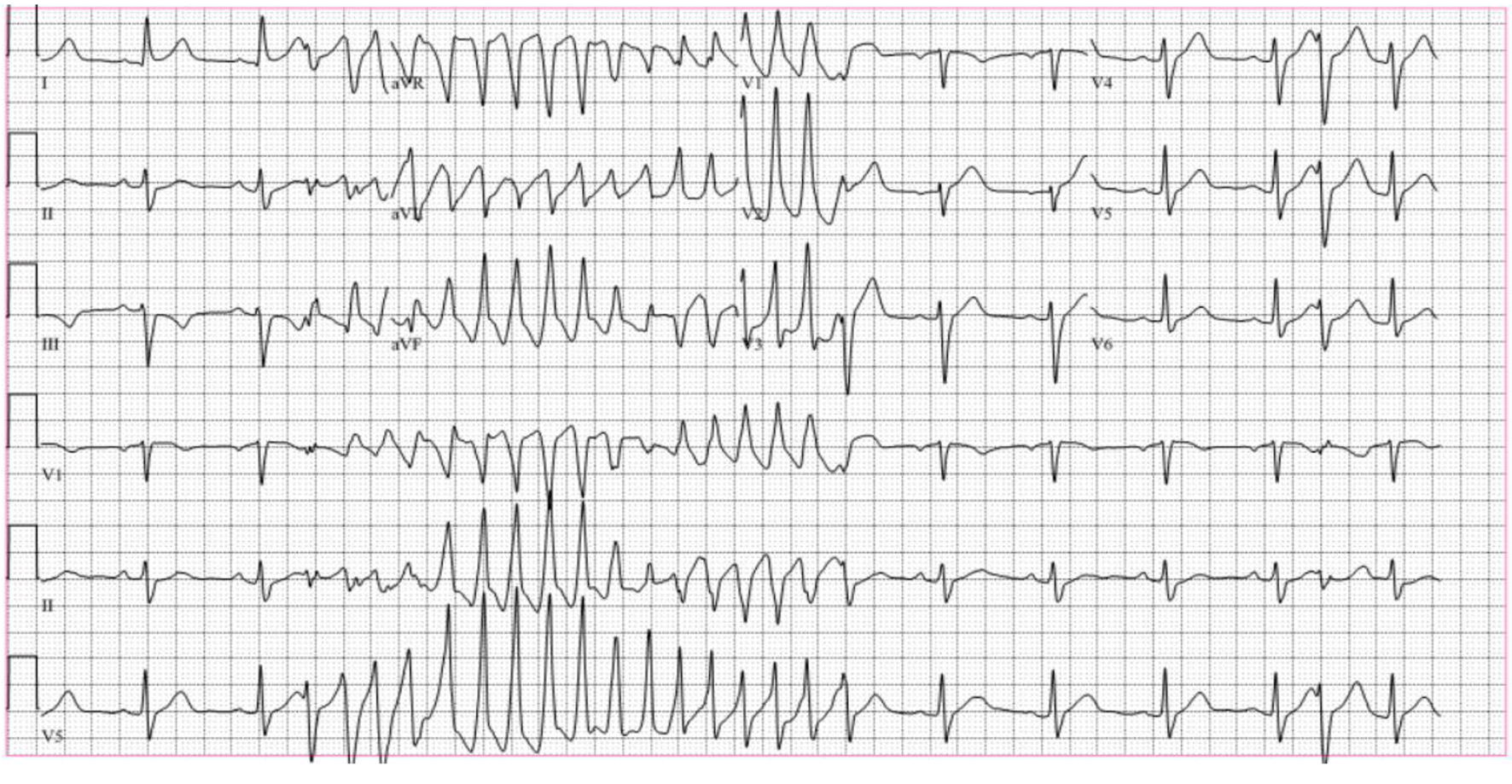
Polymorphic Ventricular Tachycardia Secondary to Subarachnoid Haemorrhage: A Rare Occurrence in the Setting of Normal QTc
Figures