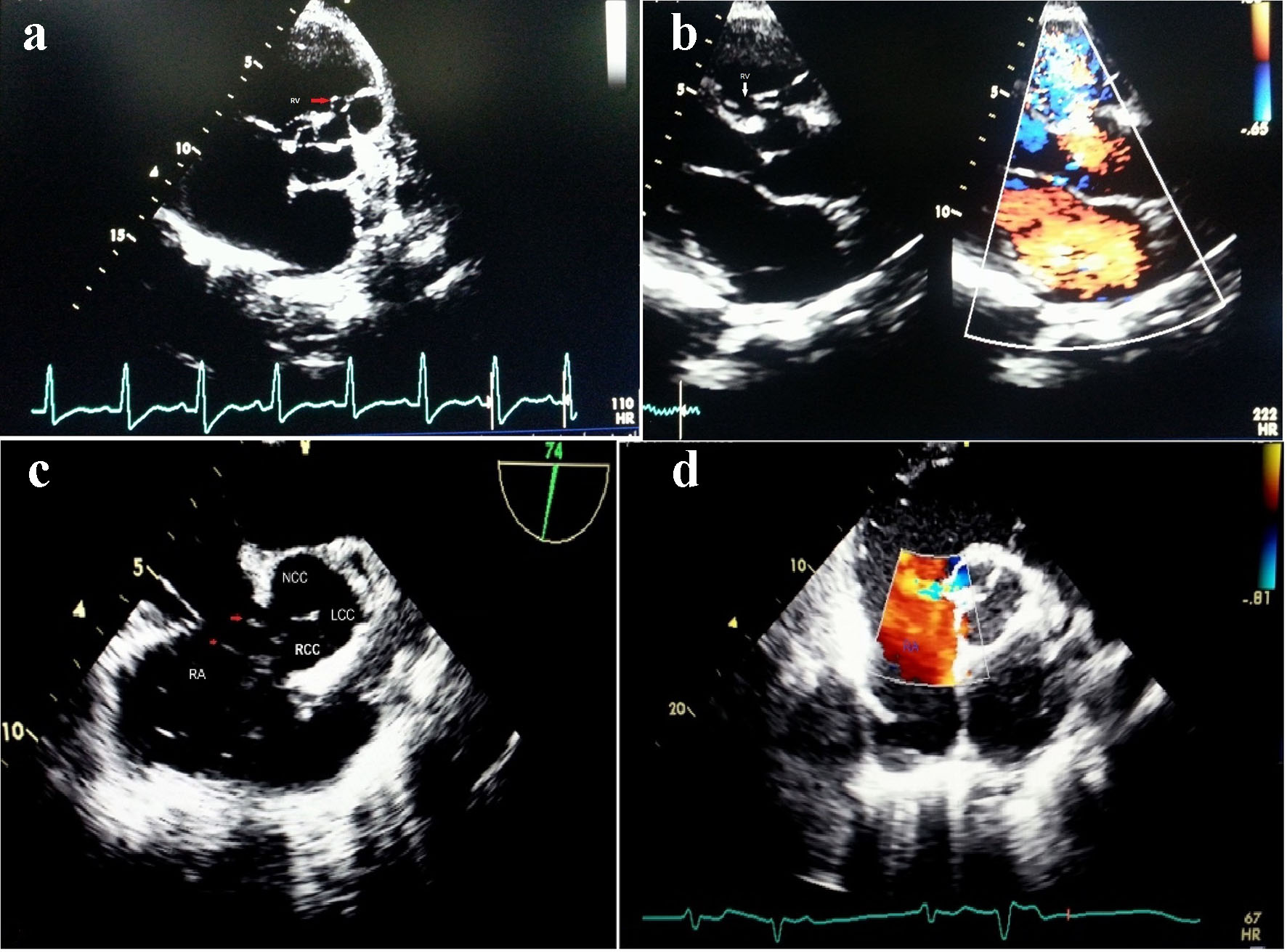
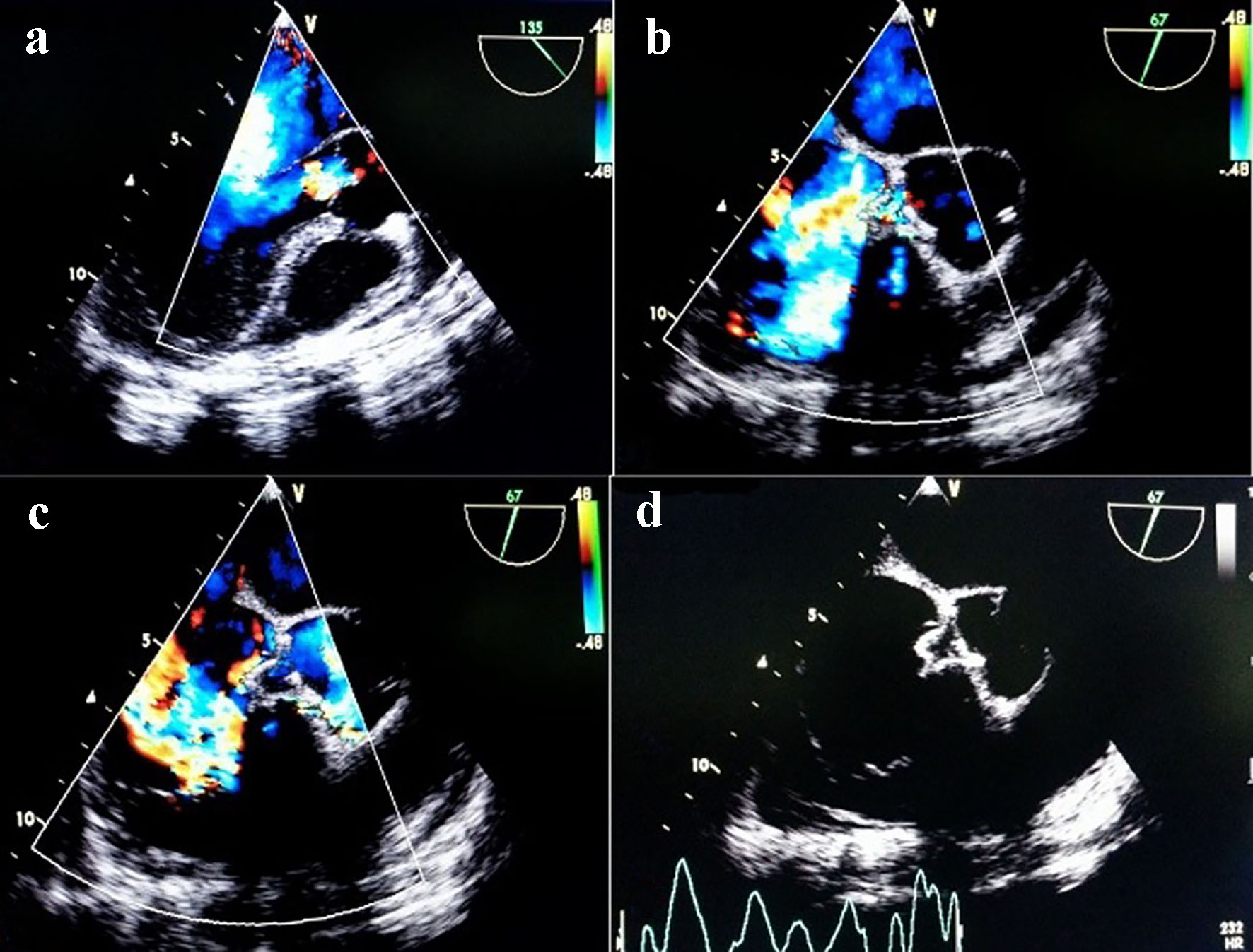
Figure 1. TTE view showing SOVA arising from right coronary cusp into right ventricle (a, b); non-coronary cusp rupturing into right atrium on TEE view (c, d).
| Cardiology Research, ISSN 1923-2829 print, 1923-2837 online, Open Access |
| Article copyright, the authors; Journal compilation copyright, Cardiol Res and Elmer Press Inc |
| Journal website https://www.cardiologyres.org |
Original Article
Volume 8, Number 4, August 2017, pages 154-160
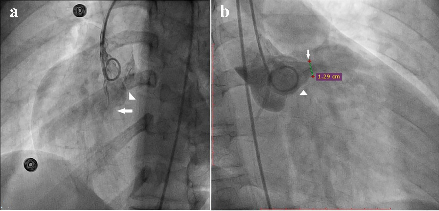
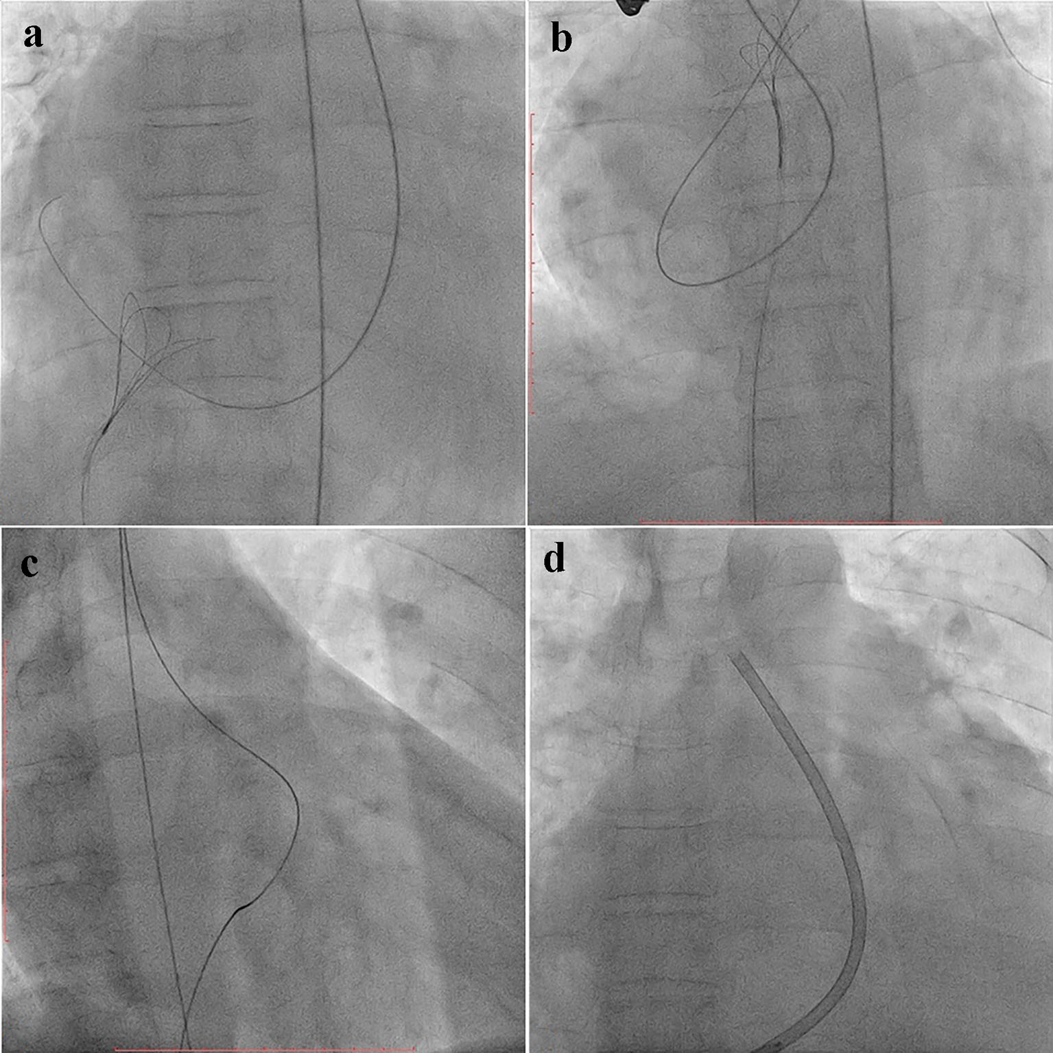
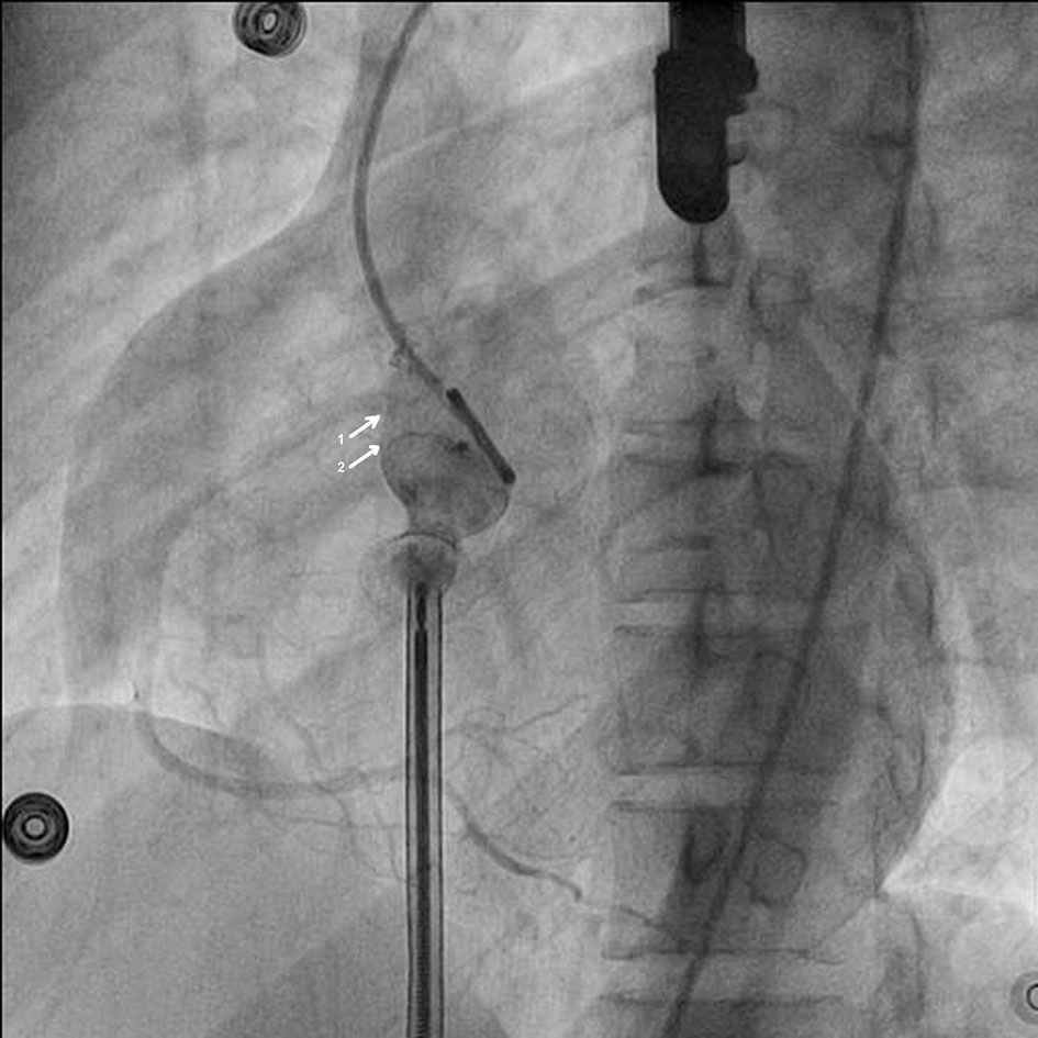
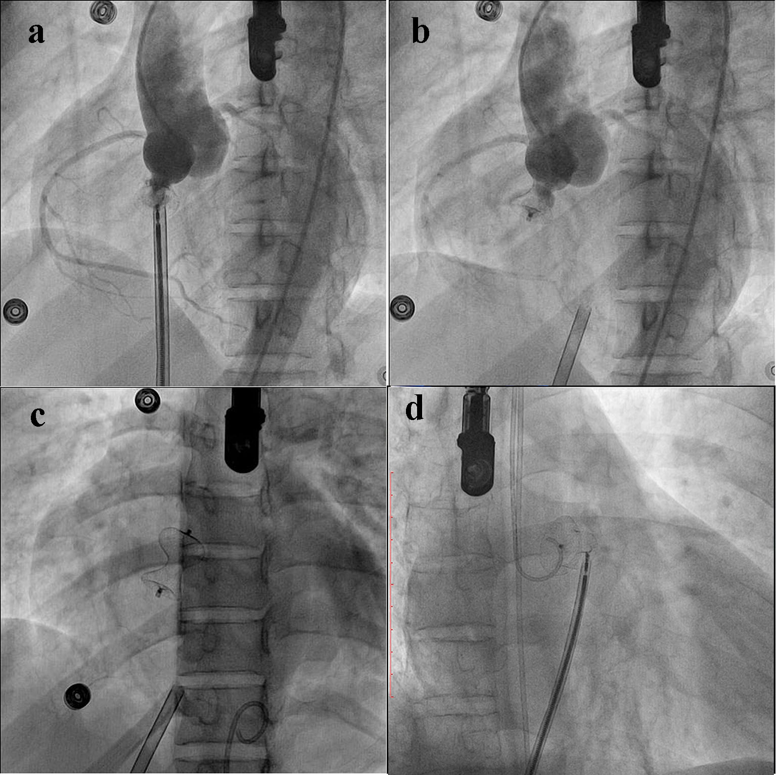
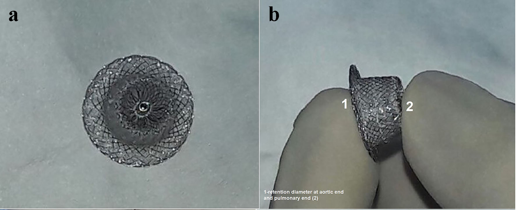
Safety and Feasibility of Transcatheter Interruption of Ruptured Sinus of Valsalva Aneurysm Using the Cocoon Duct Occluder: Immediate Results and Mid-Term Follow-Up
Figures
Table
| S. no. | Age/sex | NYHA class | Defect location | Defect size (mm) | Associated lesion | Device size | Immediate result | Follow-up (months) | Residual shunt | NYHA class |
|---|---|---|---|---|---|---|---|---|---|---|
| F: female; M: male; NYHA: New York Heart Association; RCC: right coronary cusp; RA: right atrium; NCC: non-coronary cusp; RVOT: right ventricular outflow tract; AR: aortic regurgitation; BAV: bicuspid aortic valve; TR: tricuspid regurgitation; IE: infective endocarditis; RVD: right ventricular dysfunction. | ||||||||||
| 1 | 24/M | IV | RCC-RA | 17 | Mild AR, BAV | 20/18 | Mild shunt | 13 | None | I |
| 2 | 21/F | IV | NCC-RA | 9 | None | 12/10 | No shunt | 11 | None | I |
| 3 | 19/F | III | NCC-RA | 14 | None | 18/16 | No shunt | 9 | None | I |
| 4 | 29/M | IV | RCC-RV | 10 | Severe TR, RVD | 12/10 | No shunt | 12 | None | I |
| 5 | 34/M | III | NCC-RA | 12 | Healed IE, Mild AR | 16/14 | No shunt | 15 | None | I |
| 6 | 30/M | III | NCC-RA | 16 | None | 20/18 | Trivial shunt | 12 | None | I |
| 7 | 31/F | III | NCC-RA | 14 | None | 18/16 | Trivial shunt | 22 | None | I |
| 8 | 42/M | IV | NCC-RA | 12 | Severe TR, RVD | 16/14 | No shunt | 24 | Mild TR | I |