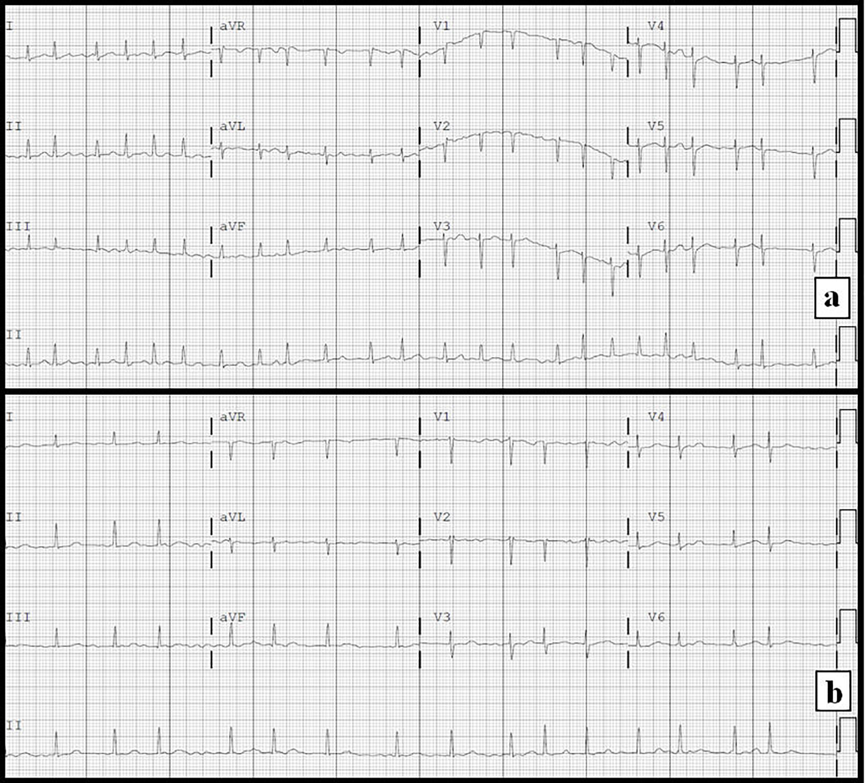
Figure 1. Electrocardiograms. (a) On admission demonstrating atrial fibrillation with rapid ventricular response. (b) Following administration of intravenous diltiazem demonstrating atrial fibrillation with a more controlled ventricular response.
| Cardiology Research, ISSN 1923-2829 print, 1923-2837 online, Open Access |
| Article copyright, the authors; Journal compilation copyright, Cardiol Res and Elmer Press Inc |
| Journal website https://www.cardiologyres.org |
Case Report
Volume 8, Number 3, June 2017, pages 134-138
Thyrotoxic Valvulopathy: Case Report and Review of the Literature
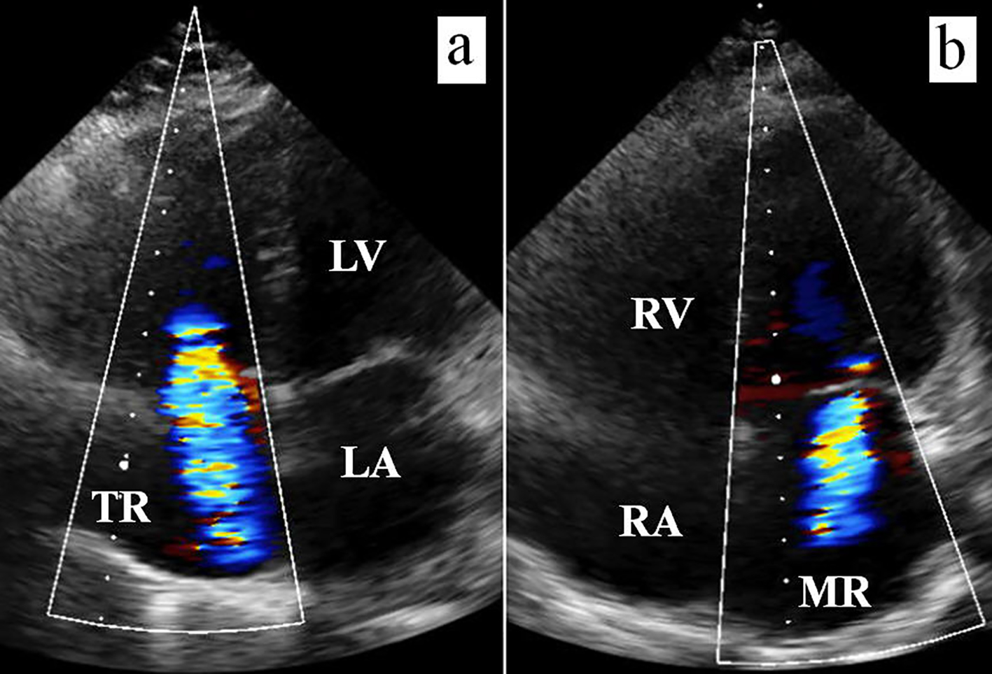
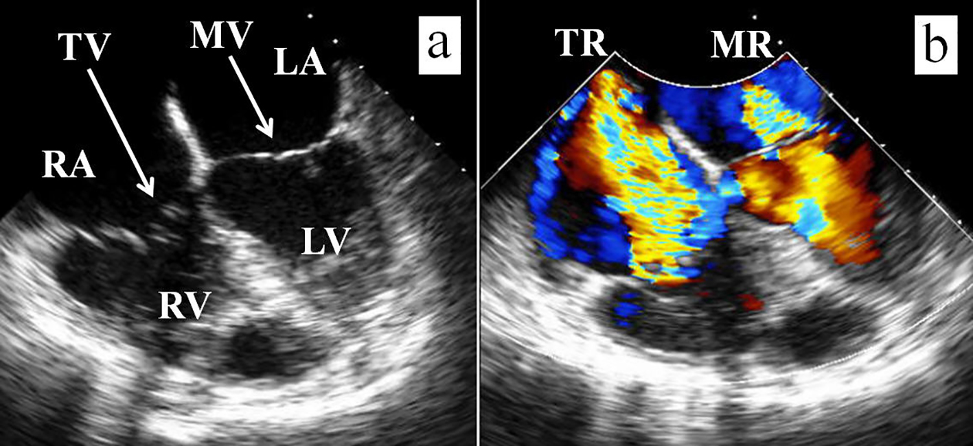
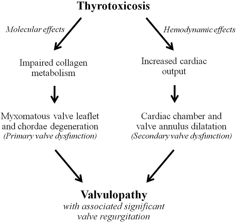
Figures