Cardiology Research, ISSN 1923-2829 print, 1923-2837 online, Open Access
Article copyright, the authors; Journal compilation copyright, Cardiol Res and Elmer Press Inc
Journal website https://www.cardiologyres.org

Case Report

Volume 8, Number 2, April 2017, pages 63-67


Cardiogenic Shock, Acute Severe Mitral Regurgitation and Complete Heart Block After Cavo-Tricuspid Isthmus Atrial Flutter Ablation

Figures

Figure 1.
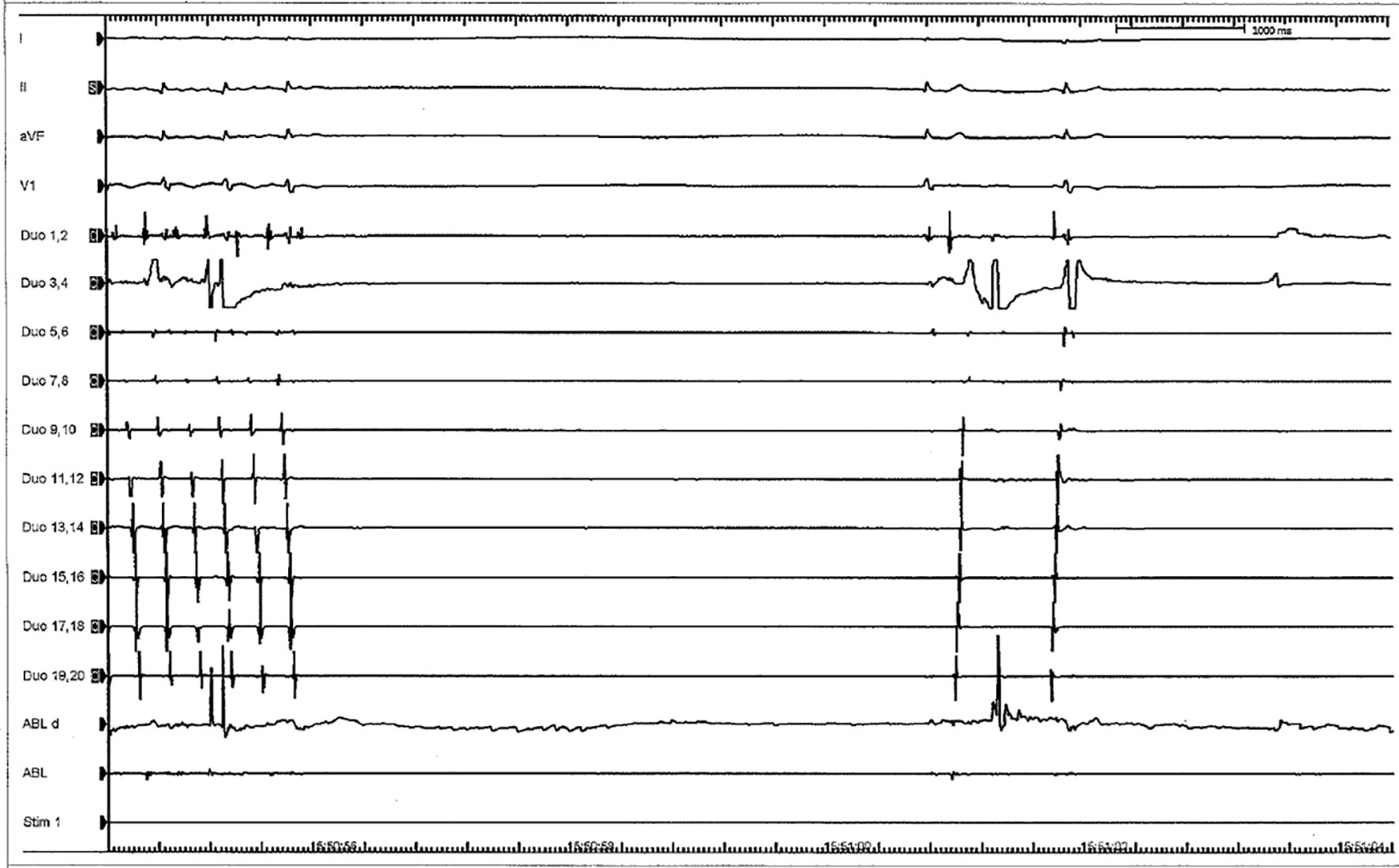
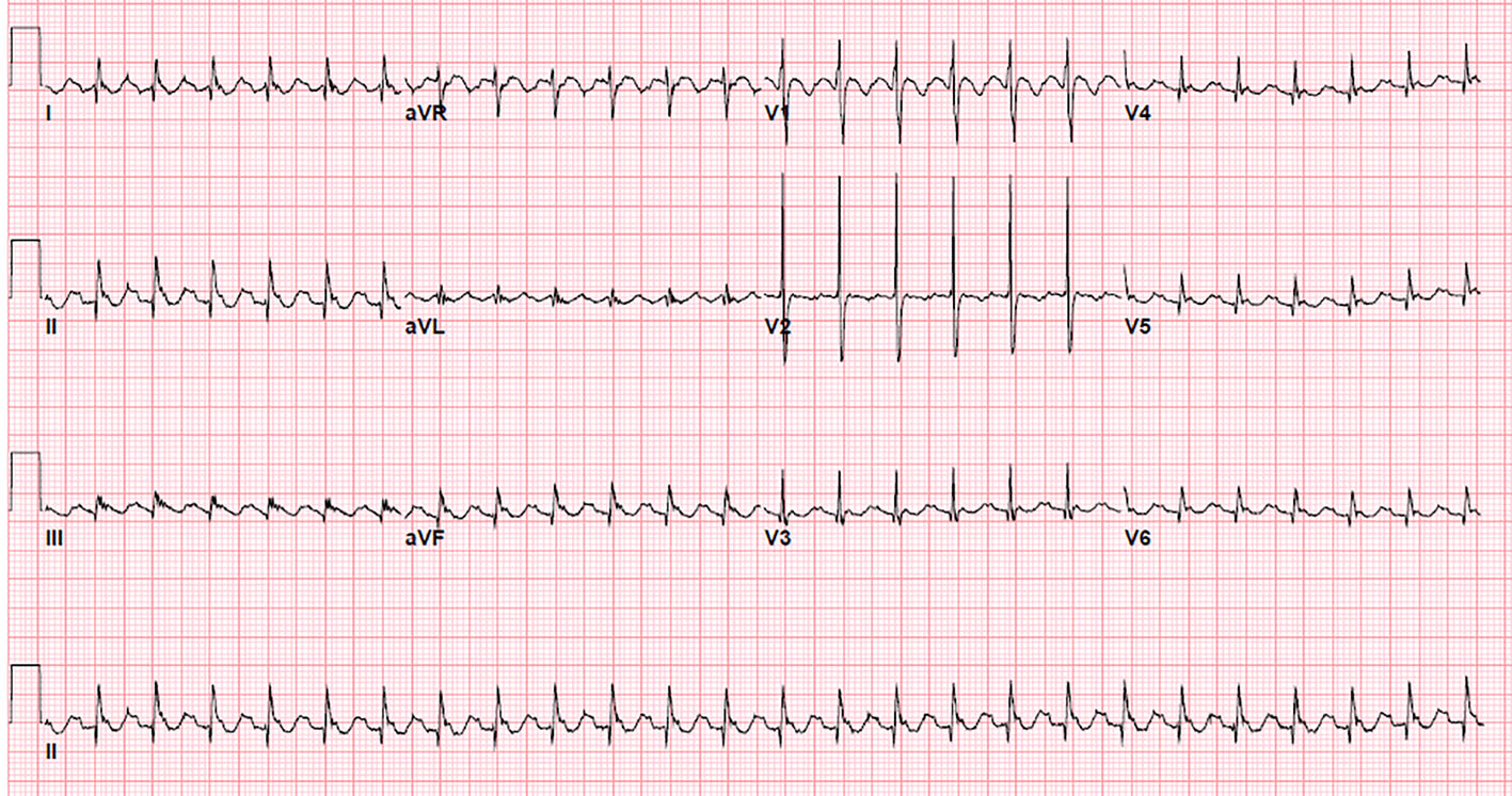
Figure 1. 12-lead surface electrogram showing saw tooth flutter waves: negative P waves in inferior leads and positive P waves in lead V1 and V2, suggestive of cavo-tricuspid isthmus dependent atrial flutter.
Figure 2.
Figure 2. Cessation of atrial flutter after radiofrequency ablation and restoration of sinus rhythm.
Figure 3.
Figure 3. Acute severe (wide open) mitral regurgitation with retracted posteromedial leaflet.
Figure 4.
Figure 4. Complete occlusion of the posterolateral branch of right coronary artery.
Figure 5.
Figure 5. After coronary stenting, flow is re-established in occluded posterolateral branch.
Figure 6.
Figure 6. Schematic drawing of the CTI and course of the RCA. The center of the RCA was tagged in three dimensions. The CTI was defined by the insertion of the IVC to the right atrium and the tricuspid valve which were also tagged. The distance of the RCA to the CTI was calculated for the anterior (ant.), inferior (inf.), and septal (sept.) parts. In addition, the deviation of the RCA from the tricuspid valve plane towards the atrial aspect was calculated. SVC: superior vena cava; CT: crista terminalis; ER: Eustachian ridge; CS: coronary sinus; AV: atrioventricular nodal branch; PDA: posterior descending artery; PLA: posterolateral artery; IVC: inferior vena cava [7].