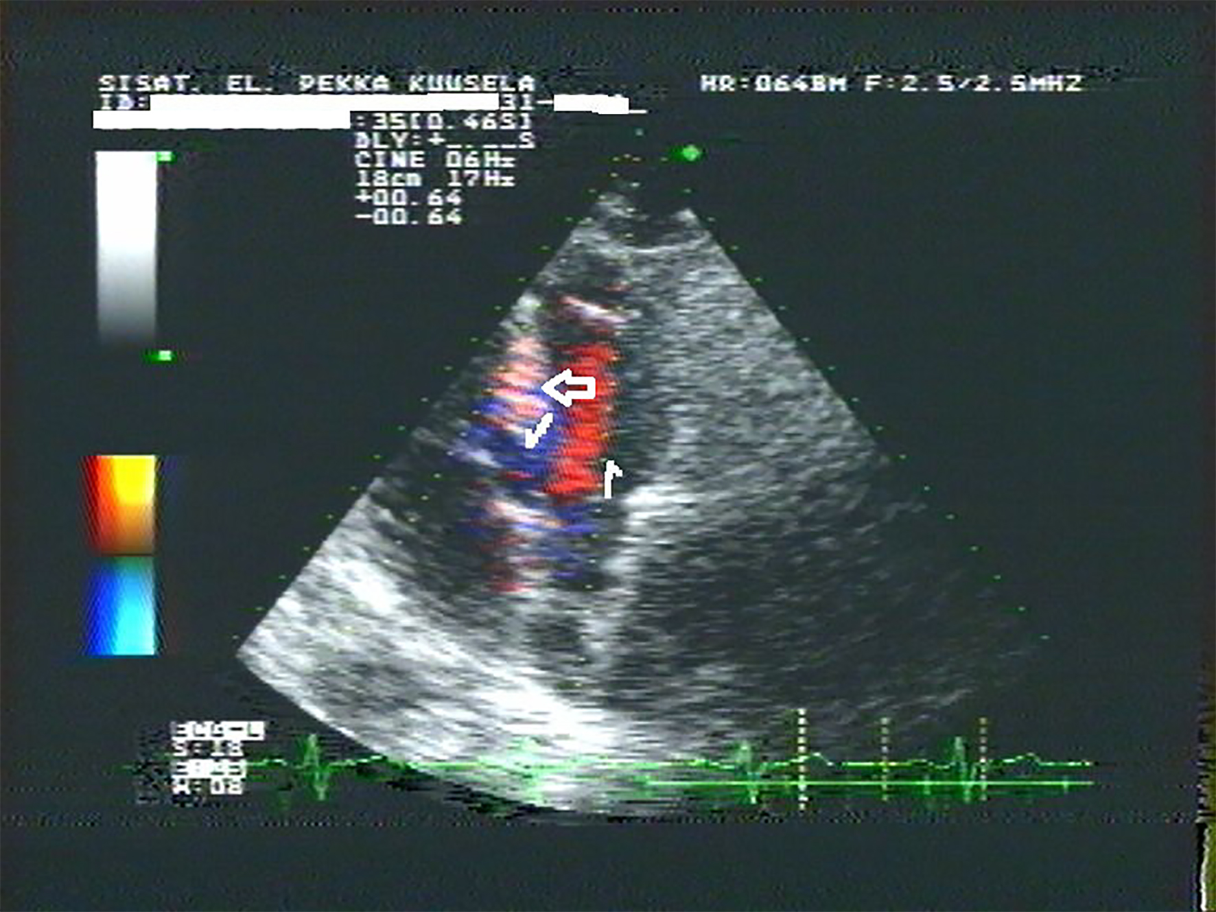
Figure 1. The right atrial filling phase was toward the apex of the RV on the right side of the septal cusp of the tricuspid valve (TV) (right arrow). The flow was again between the septum and the septal cusp of the TV toward the interventricular sinus (left arrow). The flow was from right to left through the interventricular sphincter into the LV (high arrow).
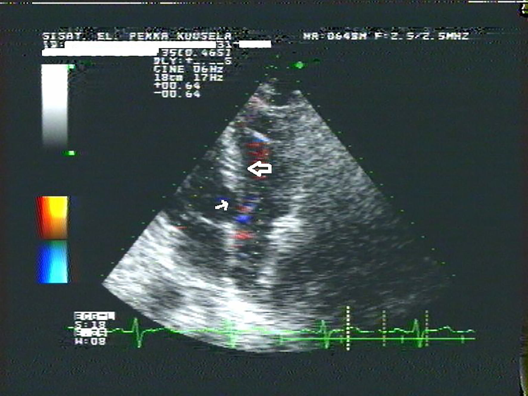
Figure 2. The end of the diastole 0.12 s later than Figure 1. The limit was visible between the right and the left muscular part of the IVS formed of the opposing fetal medial walls of the RV and the LV (low arrow). The openings of the interventricular vessel (kuuselian vessel) into the LV were visible between the interventricular sphincter in the left central muscular part of the IVS (high arrow).
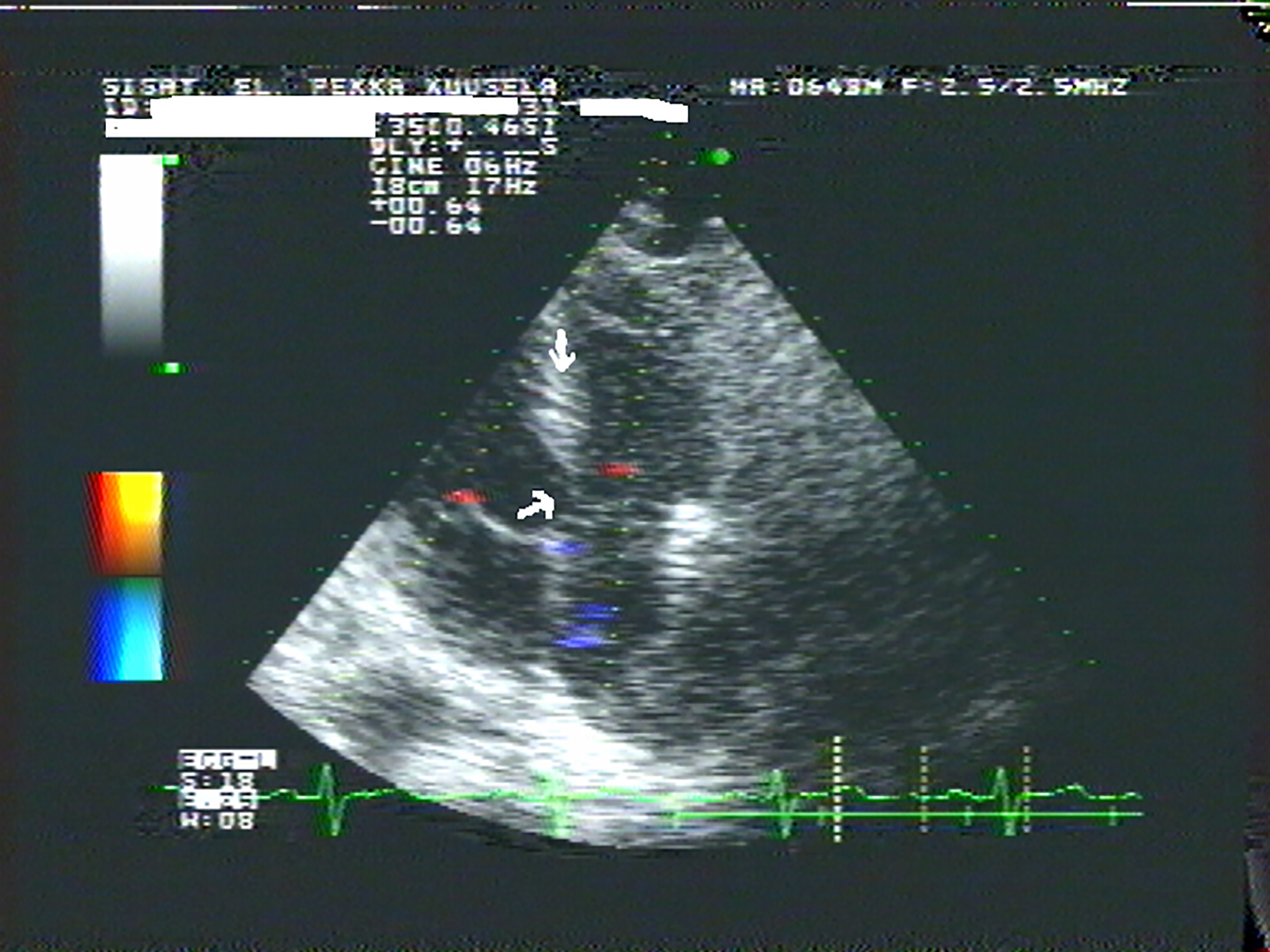
Figure 3. The limit between the right and the left muscular part of the IVS disappeared and the interventricular vessel right to the interventricular sphincter closed concordant to activation of the RV (low arrow) [1]. The interventricular sphincter exhibited closing movement concordant to activation of the middle third of the left IVS (high arrow) [1, 2].
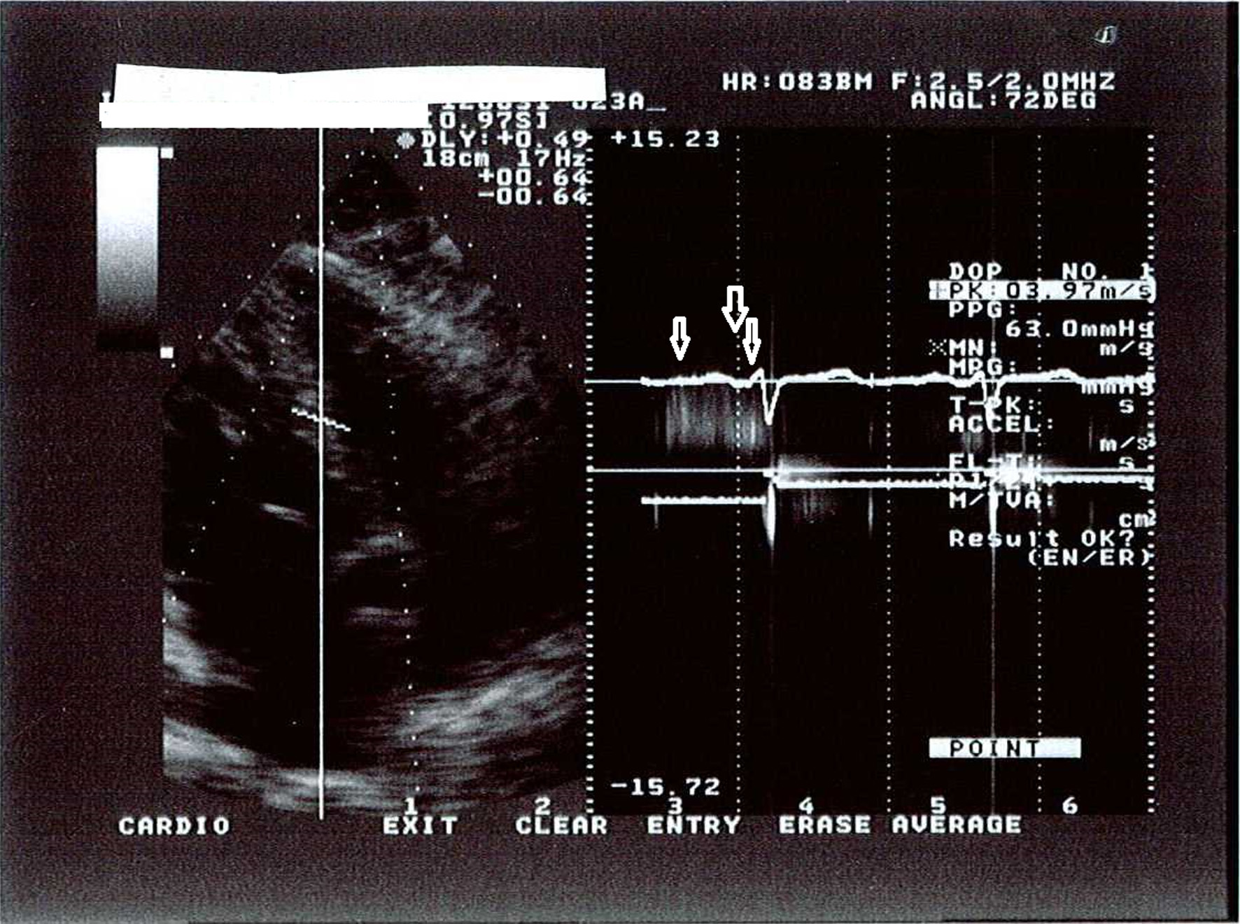
Figure 4. The CW Doppler recorded noise caused by two to three streams of blood close to each other through the openings of the interventricular vessel into the LV to demonstrate the flow at the diastole but not to measure the gradient. The gradient was not 63 mm Hg but a flow artifact. The flow through the interventricular vessel into the LV at the middle diastole suggested low left ventricular diastolic pressure (left arrow) [5]. The flow into the LV at the right atrial filling phase was concordant to the fourth heart sound (S4) (high arrow) [3, 4]. The interventricular vessel closed with a dense signal of flow as the blood ran off the interventricular vessel into the LV at the very early systole. The velocity was about 2.3 m/s and the gradient was less than 23 mm Hg (right arrow) [1, 2, 6].