Cardiology Research, ISSN 1923-2829 print, 1923-2837 online, Open Access
Article copyright, the authors; Journal compilation copyright, Cardiol Res and Elmer Press Inc
Journal website https://www.cardiologyres.org

Case Report

Volume 8, Number 2, April 2017, pages 52-56


Primary Percutaneous Coronary Intervention Angioplasty of Occluded Twin Circumflex Coronary Artery in a Patient of Acute Inferior Wall Myocardial Infarction: A Rare Anomaly

Figures

Figure 1.
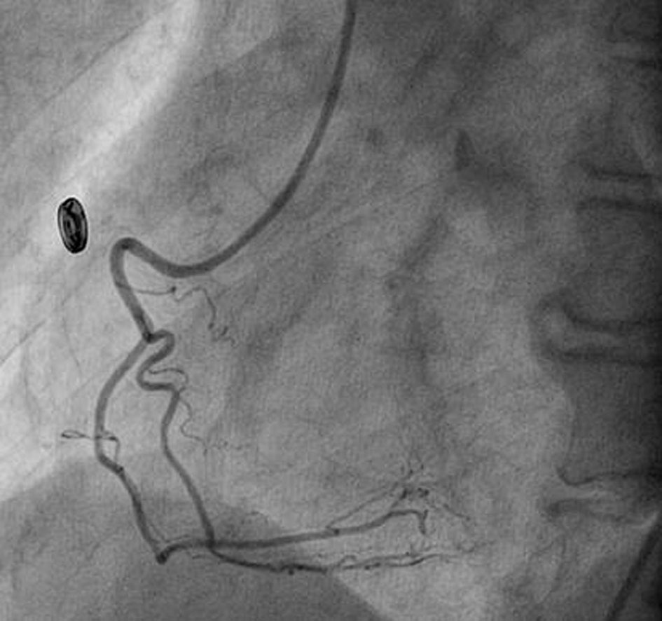
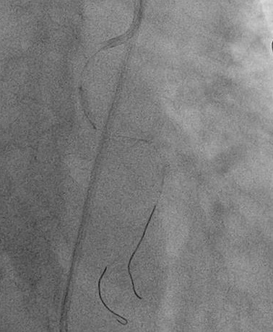
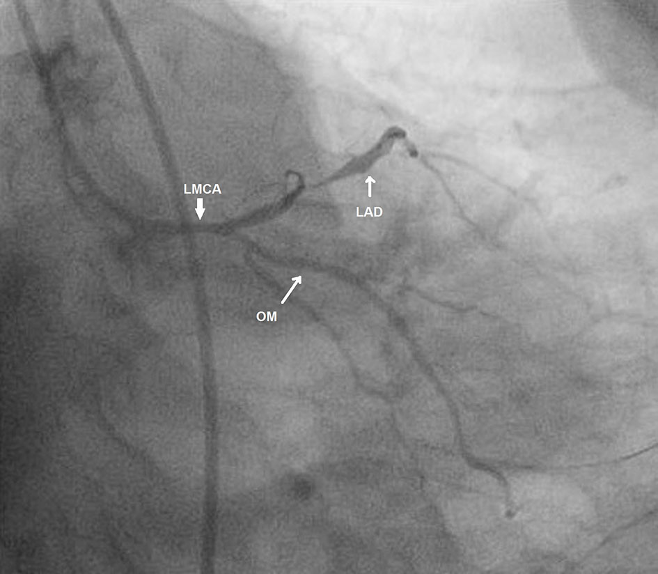
Figure 1. Left main coronary artery with left anterior descending (LAD) and obtuse marginal (OM) artery in antero-posterior caudal view.
Figure 2.
Figure 2. Normal right coronary artery.
Figure 3.
Figure 3. Anomalous LCx from RCA ostium showing critical lesion with haziness in proximal part (white arrow) in left anterior oblique view.
Figure 4.
Figure 4. Anomalous LCx from RCA ostium showing critical lesion with haziness in proximal part (white arrow) in left anterior oblique with cranial tilt.
Figure 5.
Figure 5. Improvization of Judkins right catheter by straightening its tip.
Figure 6.
Figure 6. Cannulated LCx with help of buddy wire and lesion being pre-dilated with 2 × 10 mm Minitrak balloon.
Figure 7.
Figure 7. Lesion being stented by 2.75 × 33 mm Xience Prime drug-eluting stent (Abott, USA).
Figure 8.
Figure 8. Stent being post-dilated by 2.75 × 10 mm Minitrak non-compliant balloon.
Figure 9.
Figure 9. TIMI 3 flow in LCx after dilatation.