Figures
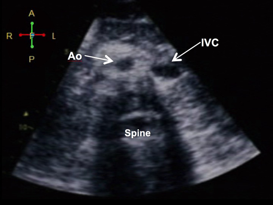
Figure 1. Demonstration of situs inversus. (A) Subcostal view showing the aorta (Ao) and the inferior vena cava (IVC) in relation to the spine: the aorta (more pulsatile) is at the right while the IVC (collapsible) is on the left (visceral situs inversus). The liver is on the left and the spleen and stomach are on the left (not shown).
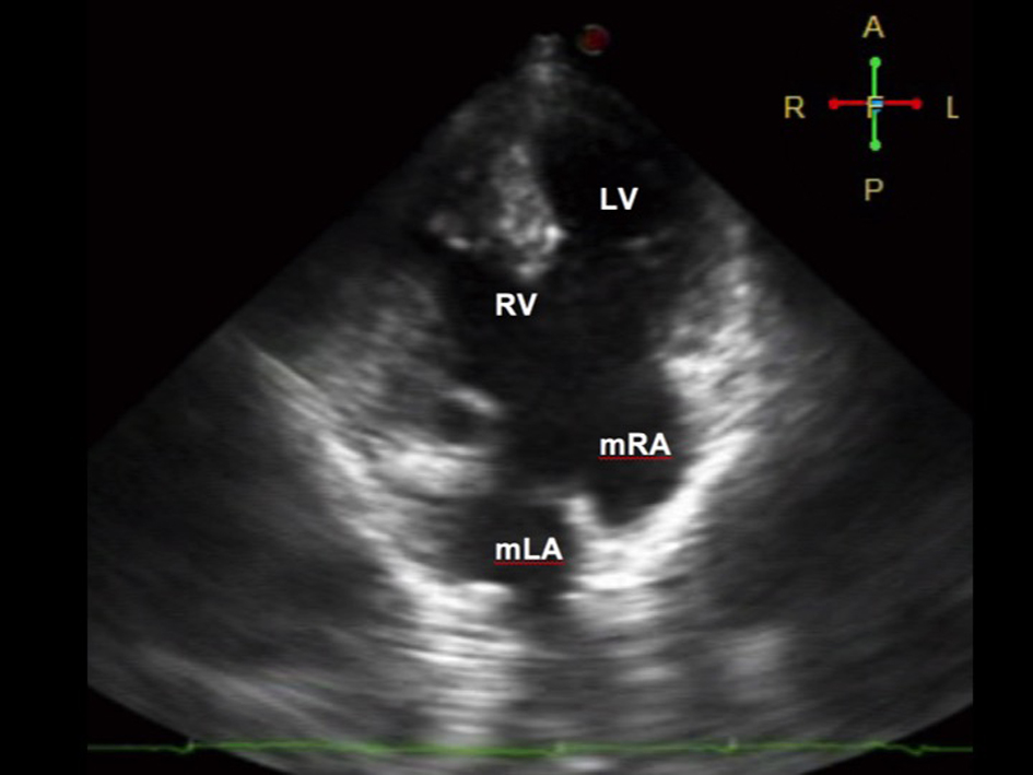
Figure 2. Four-chamber view on 2D echocardiography during diastole showing a complete atrioventricular (AV) septal defect Rastelli type A. There is a common AV valve, which makes it difficult to use the valves to identify the ventricles. Note that the moderator band is seen on the ventricle of the right (morphologic RV). The morphologic right atrium (mRA) drains into the left ventricle, while the morphologic left atrium (mLA) drains into the right ventricle, suggestive of atrioventricular discordance. The RV (systemic chamber) is more hypertrophied than the LV.
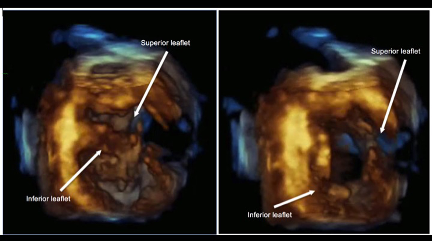
Figure 3. 3D echocardiography demonstrating a common AV valve during systole (left) and diastole (right) taken at short axis view with its corresponding M-mode. No significant regurgitation was noted on color flow Doppler.
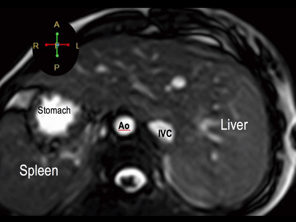
Figure 4. Visceral situs inversus seen on cardiac MRI. Axial cut at the level of the left-sided liver and right-sided spleen and stomach. The aorta (Ao) is on the right while the inferior vena cava (IVC) is on the left.
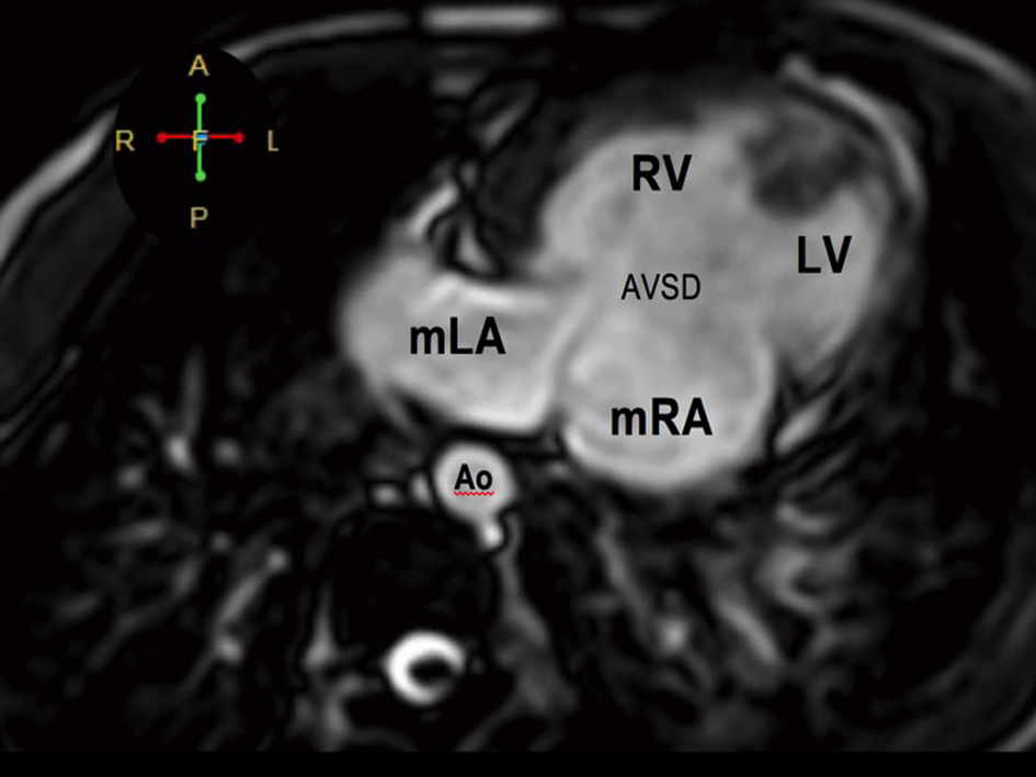
Figure 5. Atrioventricular discordance on cardiac MRI. The morphologic LA drains into the morphologic RV while the morphologic RA drains into the morphologic LV. There is a complete atrioventricular septal defect (AVSD).
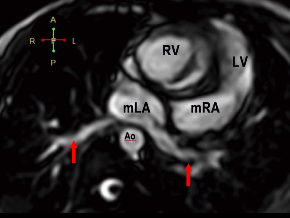
Figure 6. The pulmonary veins drain into the morphologic left atrium located on the right side (atrial situs inversus).
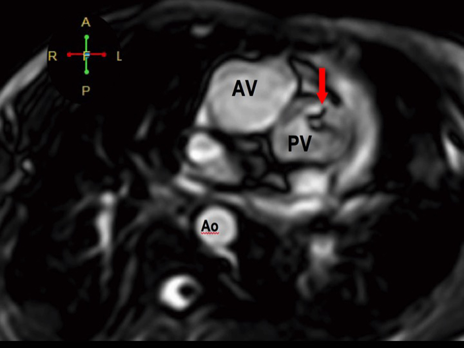
Figure 7. The RV eventually drains into the malposed aorta (Ao) and main pulmonary artery (MPA), indicative of DORV with malposition. There is also note of pulmonic stenosis (arrow).
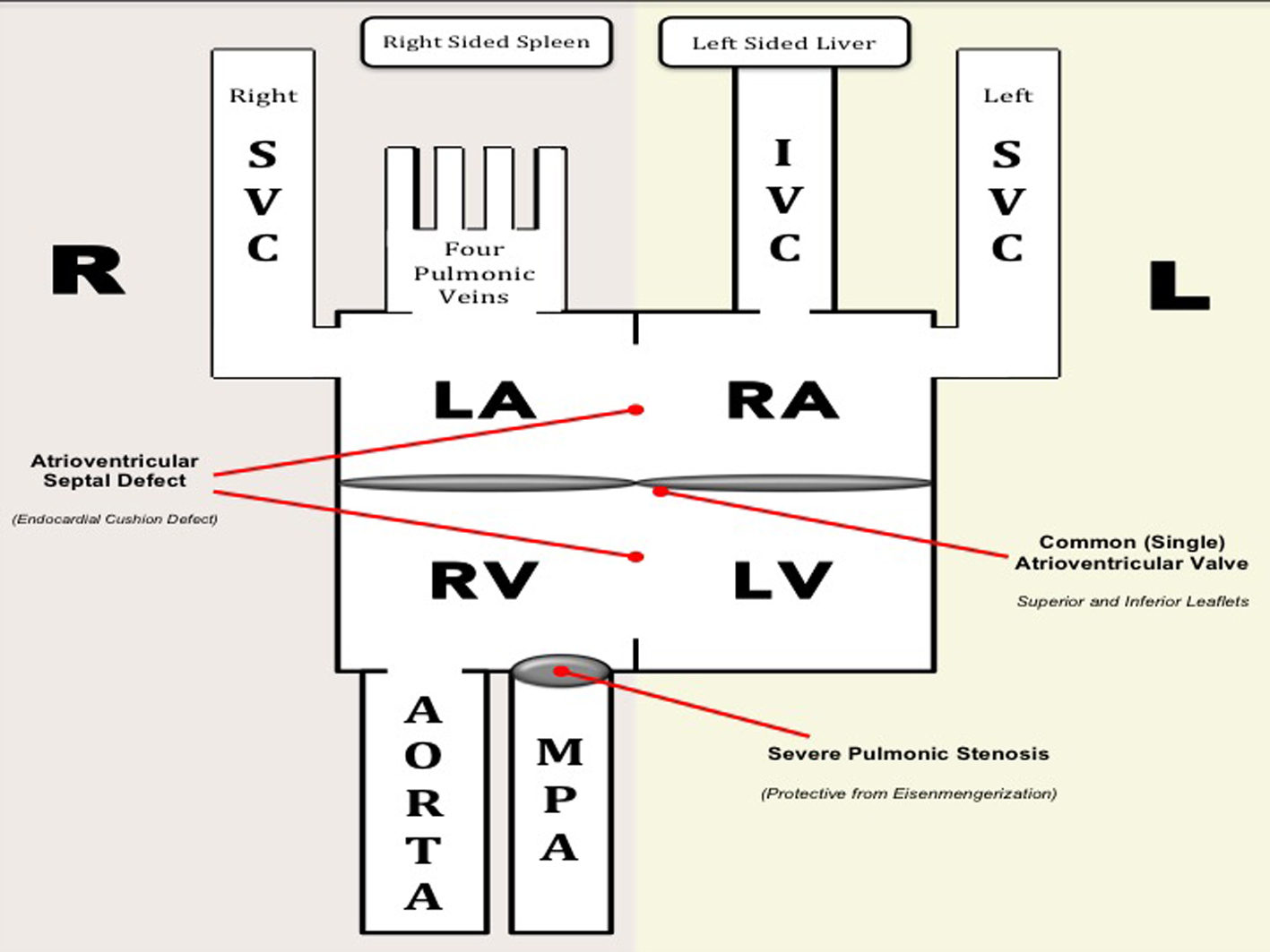
Figure 8. Hemodynamics of our patient. Deoxygenated blood from the IVC and left SVC enter the morphologic RA and blood from the right SVC enter the morphologic LA. Oxygenated blood from the four pulmonary veins drain into the morphologic LA. The morphologic RA and LA are separated by a short segment of the interatrial septum. Functionally, deoxygenated blood from the three vena cavae and oxygenated blood from the pulmonary veins enter a common (single) atrium. Mixed blood from the atria drain across a common AV valve into a functionally univentricle. Anatomically, both ventricles are separated from each other by a short interventricular septum. Mixed blood from the functional univentricle drain into the aorta and main pulmonary artery, both of which arise from the RV (DORV). Note that there is a severe pulmonic stenosis which is protective from the development of pulmonary hypertension (i.e. Eisenmengerization). Diagram on the right represents the normal anatomy.