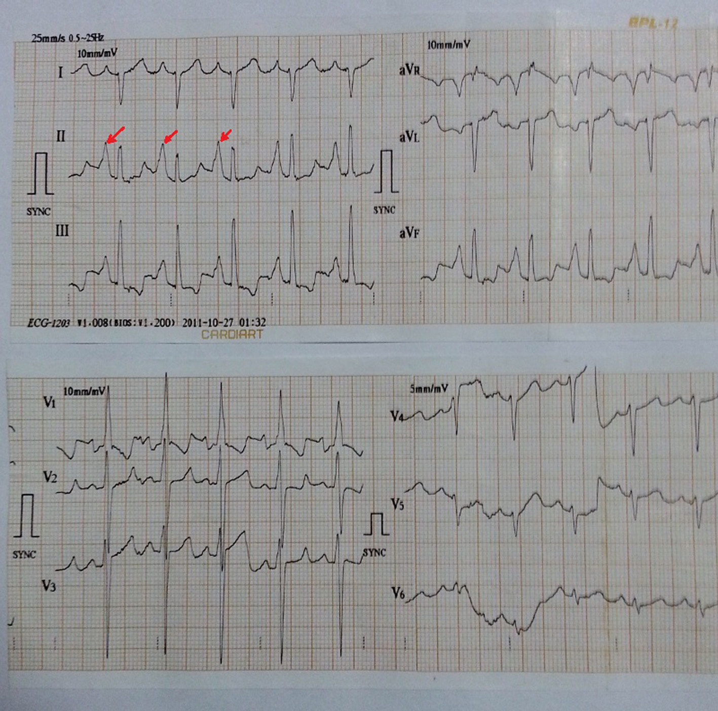
Figure 1. In lead II, III and aVF, P-wave was tall and peaked with maximum P-wave amplitude > 7 mm and larger than QRS complex in lead II. P-wave axis is +82°. Precordial leads also show right ventricular hypertrophy with strain.
| Cardiology Research, ISSN 1923-2829 print, 1923-2837 online, Open Access |
| Article copyright, the authors; Journal compilation copyright, Cardiol Res and Elmer Press Inc |
| Journal website https://www.cardiologyres.org |
Case Report
Volume 6, Number 4-5, October 2015, pages 336-338
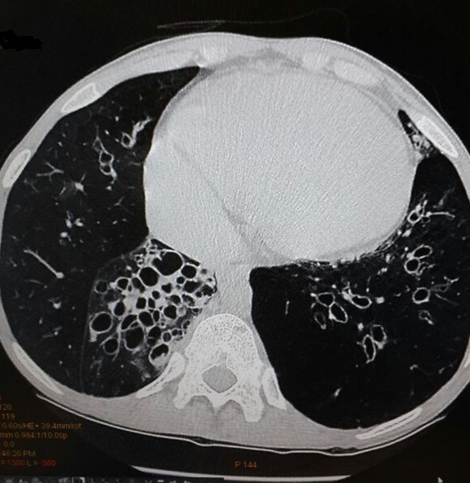
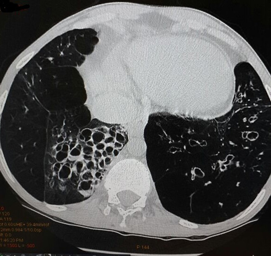
Rare Electrocardiographic Manifestation of Cystic Bronchiectasis in a 34-Year-Old Male
Figures