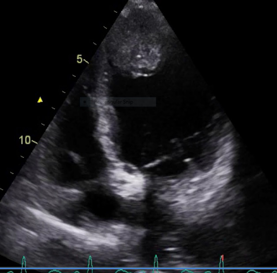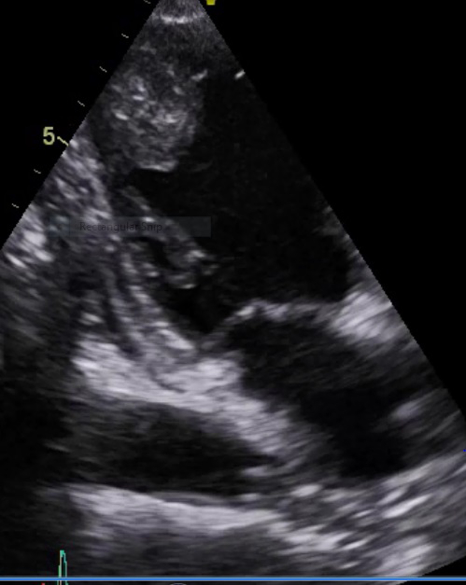
Figure 1. Apical four-chamber view showing heterogenous echogenic irregular vascular mass lesion in the endocardium with normal apical (left ventricular) mobility.
| Cardiology Research, ISSN 1923-2829 print, 1923-2837 online, Open Access |
| Article copyright, the authors; Journal compilation copyright, Cardiol Res and Elmer Press Inc |
| Journal website https://www.cardiologyres.org |
Case Report
Volume 6, Number 4-5, October 2015, pages 329-331
Squamous Cell Carcinoma of Lung Atypically Involving Heart: A Case Report With Literature Review
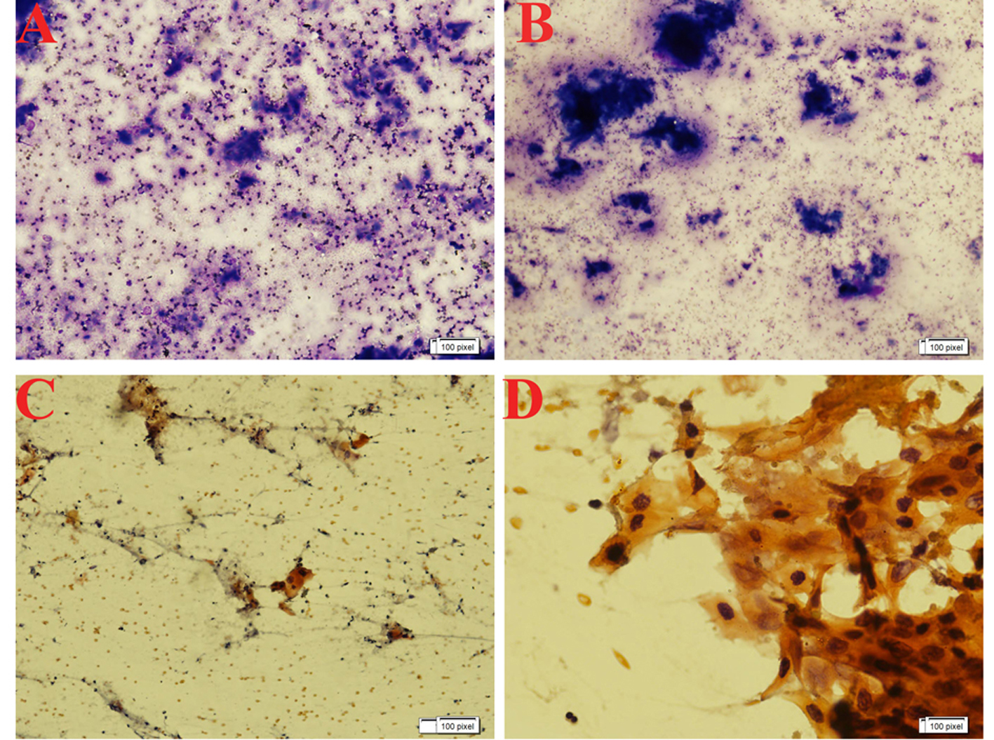
Figures