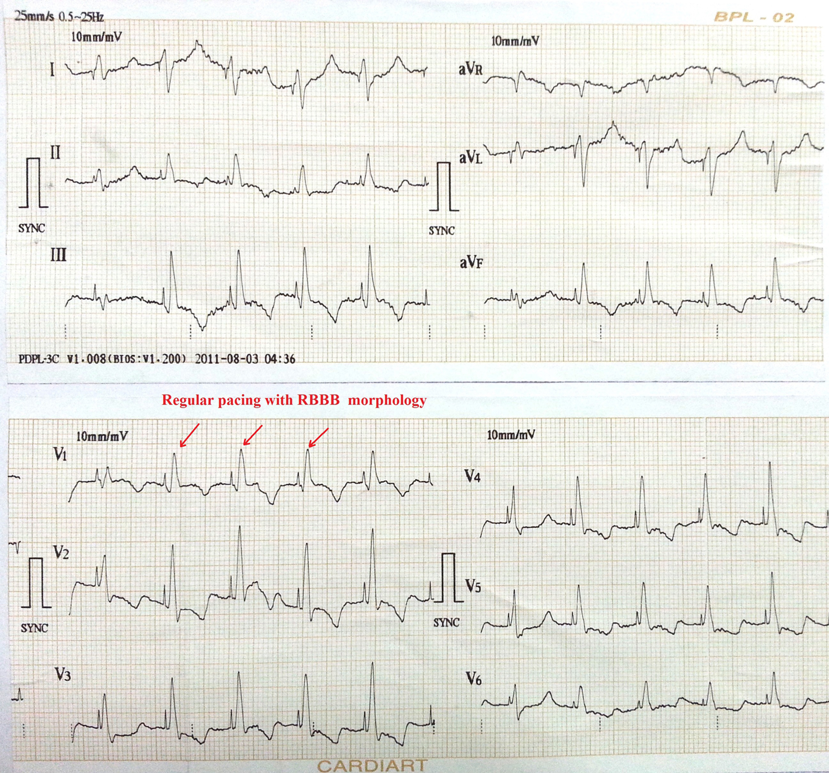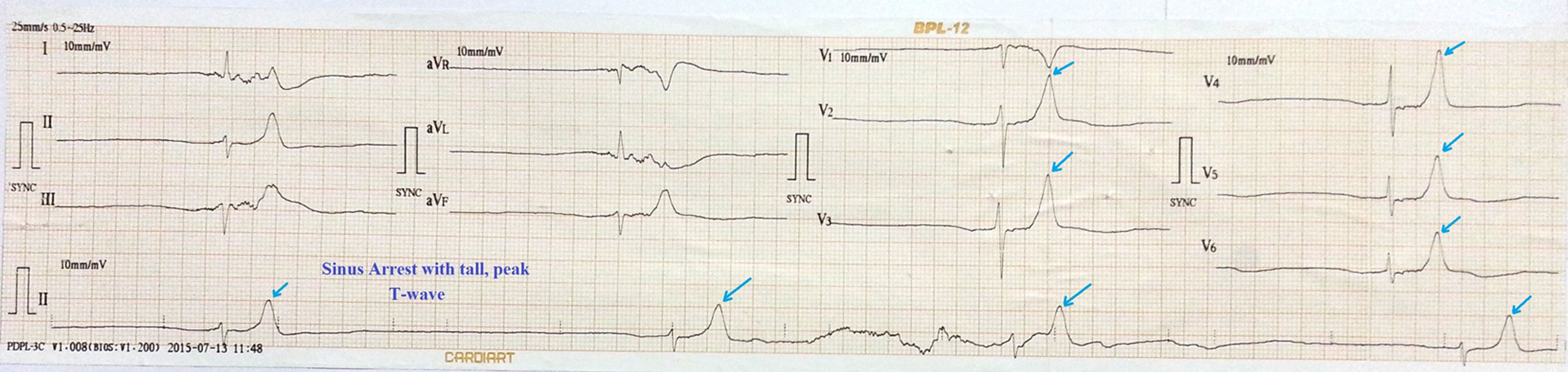
Figure 1. ECG showing sinus arrest, tall peaked T wave with heart rate of 16 bpm.
| Cardiology Research, ISSN 1923-2829 print, 1923-2837 online, Open Access |
| Article copyright, the authors; Journal compilation copyright, Cardiol Res and Elmer Press Inc |
| Journal website https://www.cardiologyres.org |
Case Report
Volume 6, Number 4-5, October 2015, pages 324-328
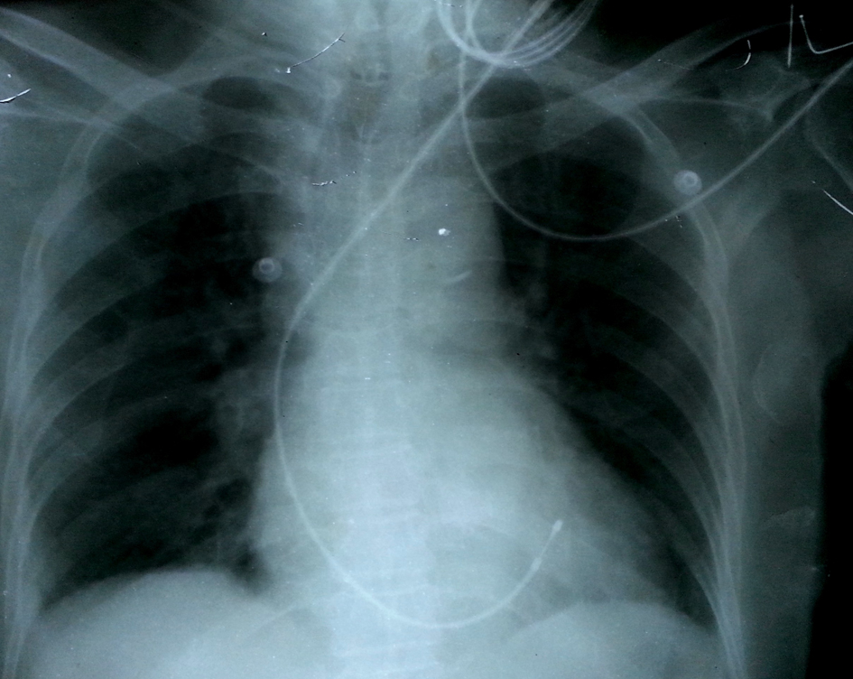
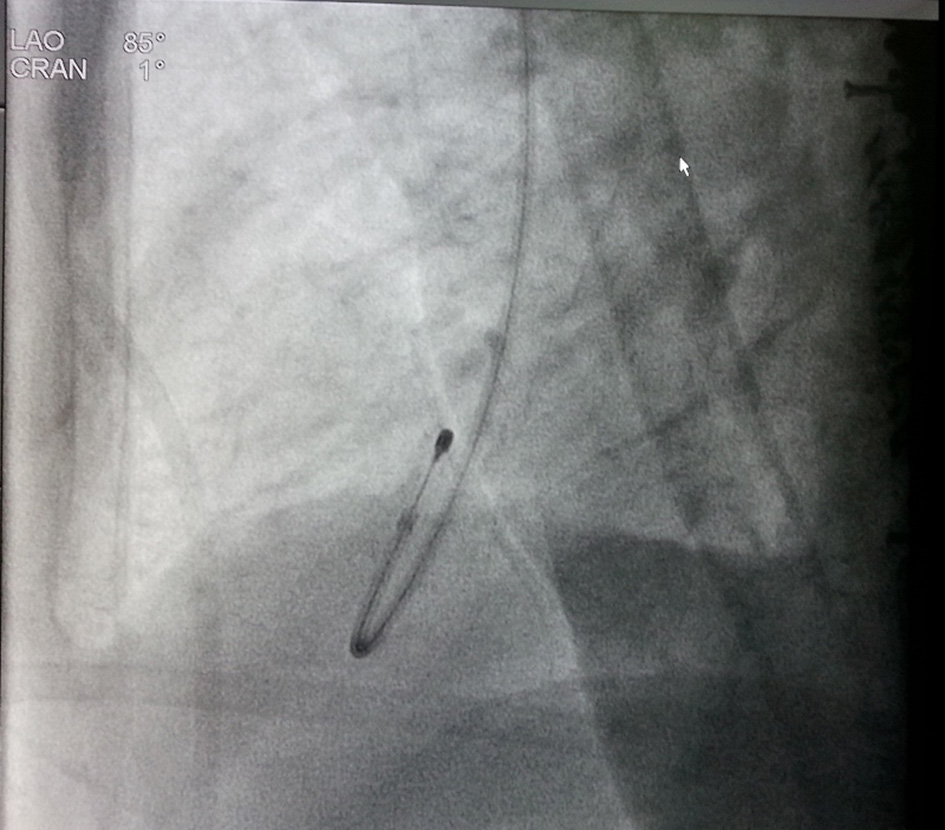
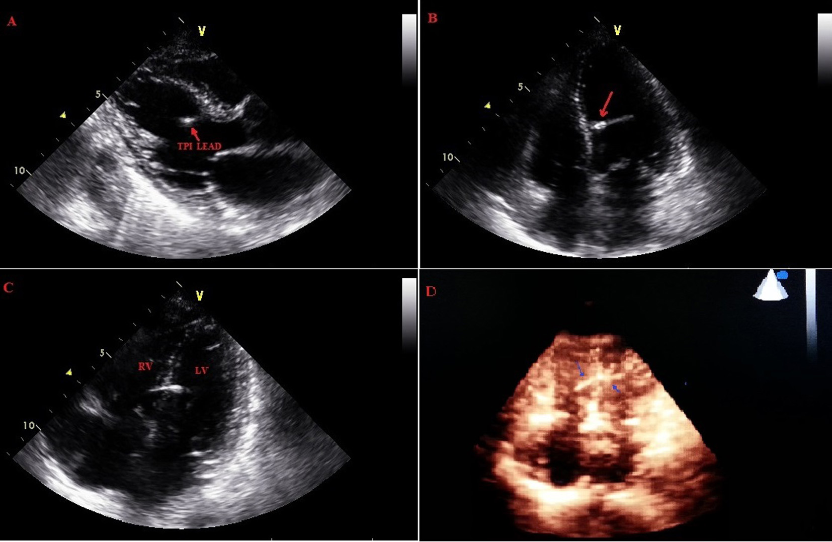
An Unconventional Route of Left Ventricular Pacing
Figures