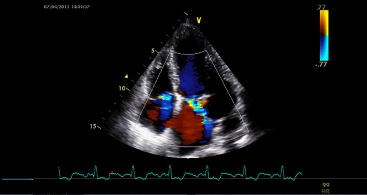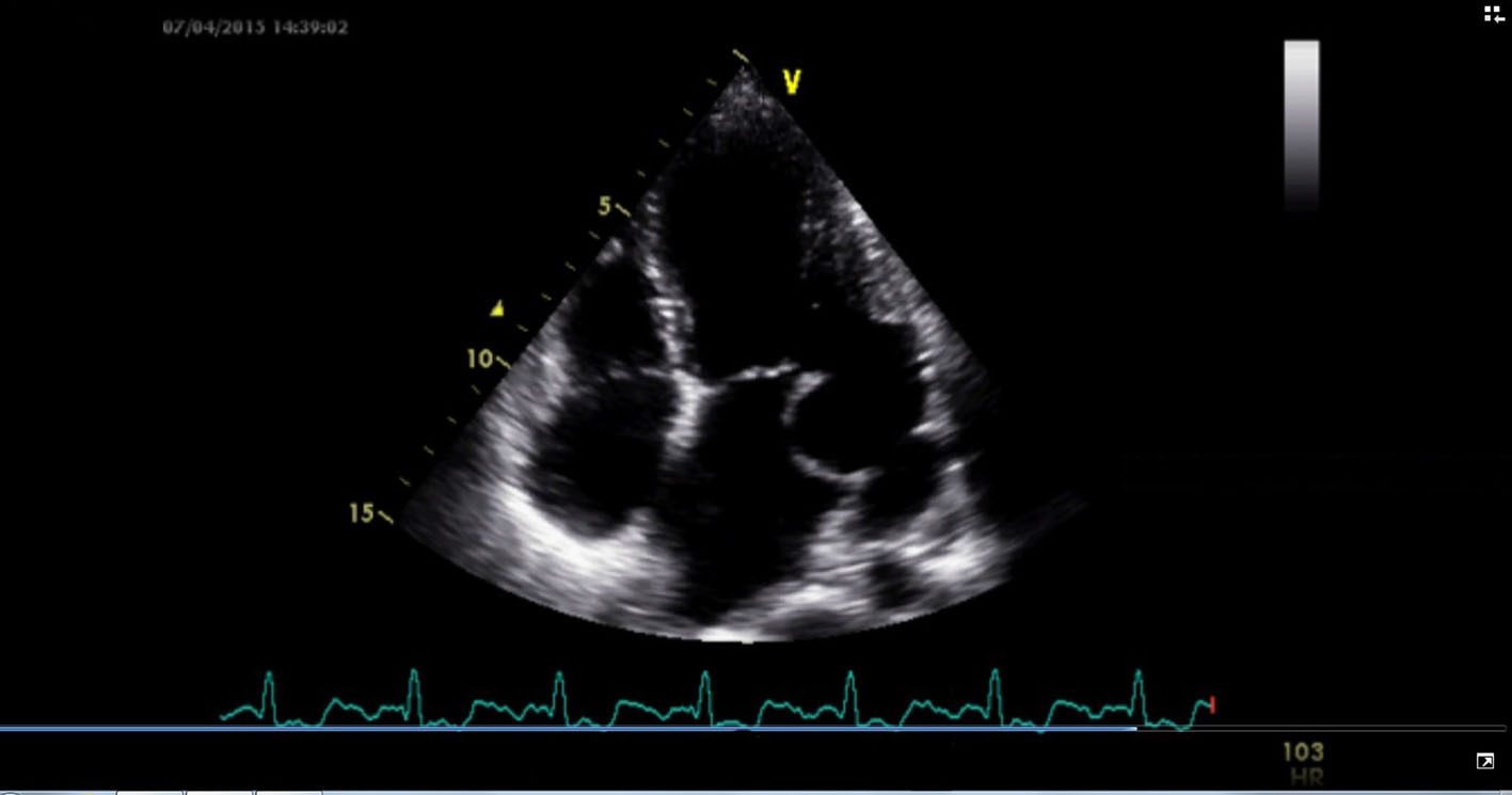
Figure 1. Transthoracic echocardiography in apical four-chamber view showing submitral aneurysm in the posterolateral wall of the left ventricle.
| Cardiology Research, ISSN 1923-2829 print, 1923-2837 online, Open Access |
| Article copyright, the authors; Journal compilation copyright, Cardiol Res and Elmer Press Inc |
| Journal website https://www.cardiologyres.org |
Case Report
Volume 6, Number 3, June 2015, pages 297-299
Subvalvar Mitral Aneurysm: A Rare Cause of Mitral Leak
Figures