Cardiology Research, ISSN 1923-2829 print, 1923-2837 online, Open Access
Article copyright, the authors; Journal compilation copyright, Cardiol Res and Elmer Press Inc
Journal website https://www.cardiologyres.org

Case Report

Volume 6, Number 3, June 2015, pages 292-296


Elusive Cardiac Angiosarcoma in a Young Pregnant Female: Rare Presentation With Fatal Outcome

Figures

Figure 1.
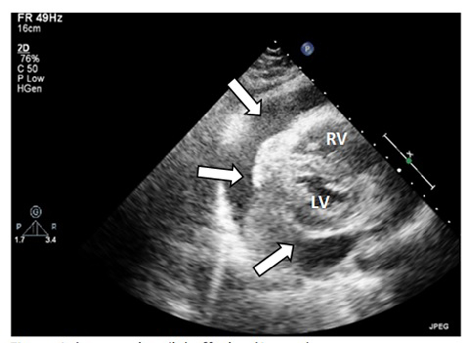
Figure 1. Large pericardial effusion (arrow). RV: right ventricle; LV: left ventricle.
Figure 2.
Figure 2. Transesophageal echocardiogram (TOE): transverse view of the right chambers. There is a large firm infiltrating mass (M) in the right atrium (RA), with a propagation (arrow) within the right atrial cavity. RA: right atrium; TV: tricuspide valve; RV: right ventricle.
Figure 3.
Figure 3. Cardiac MRI revealing large irregular tumor (T) occupying most of the RA, with numerous bilateral lung metastases (LM). RV: right ventricle; LA: left atrium; LV: left ventricle.
Figure 4.
Figure 4. Liver biopsy showing (A) photomicrograph of moderate to poorly differentiated angiosarcoma with dense clumps of plump and spindle shaped, anaplastic cells focally showing slit-like vascular lumens (original magnification, × 200); (B) entrapped hepatocytes (arrows) within the tumor (original magnification, × 400); (C) immunohistochemical staining for the endothelial marker CD34 demonstrating the endothelial origin of the neoplastic cells.