Cardiology Research, ISSN 1923-2829 print, 1923-2837 online, Open Access
Article copyright, the authors; Journal compilation copyright, Cardiol Res and Elmer Press Inc
Journal website https://www.cardiologyres.org

Case Report

Volume 6, Number 3, June 2015, pages 289-291


ALCAPA in an Octogenarian Woman: An Enigma

Figures

Figure 1.
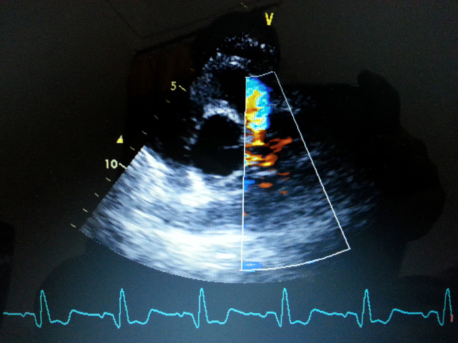
Figure 1. Transthoracic echo-parasternal short-axis view showing presence of continuous flow entering the pulmonary trunk.
Figure 2.
Figure 2. RAO (right anterior oblique) view of dilated and tortuous right coronary artery.
Figure 3.
Figure 3. RAO (right anterior oblique) view of late phase showing abundant intercoronary anastomoses (Rentrop grade 3 intercoronary collateral) communicating with left coronary artery.
Figure 4.
Figure 4. RAO (right anterior oblique) view of LAD and LCX arteries opacifying the LMCA and its origin from the main pulmonary artery.
Figure 5.
Figure 5. Reconstructed 3D CT angiogram showing dilated and tortuous right coronary artery with abundant intercoronary anastomoses communicating with left coronary artery and absence of its origin from aorta.